2026 Author: Priscilla Miln | miln@babymagazinclub.com. Last modified: 2025-01-22 17:55:18
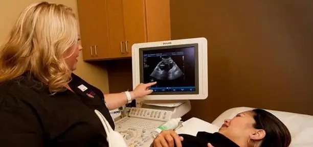
With the onset of pregnancy, a woman begins to worry about many questions. Every expectant mother wishes her baby a normal formation and development. In the early stages, there may be risks of developing certain diseases of the embryo. To study the condition of the baby, doctors prescribe screening for the 1st trimester. The norms for ultrasound (a photo of the examination is usually attached) a woman can find out from a specialist who observes her.
What is perinatal screening?
Perinatal screening involves a study of a pregnant woman, which allows to identify various malformations of the child at the stage of intrauterine development. This method includes two types of examination: a biochemical blood test and an ultrasound examination.

The optimal period for conducting such a survey has been determined - this is a period from ten weeks and six days to thirteen weeks and six days. There is a certain norm for screening ultrasound of the 1st trimester, with which the results of the examination of a pregnant woman are compared. The main task of ultrasound at this time is to determine serious malformations of the embryo and to identifymarkers of chromosomal abnormalities.
The main anomalies are:
- TVP size - the thickness of the space of the collar zone;
- underdevelopment or absence of nasal bones.
Ultrasound during pregnancy reveals signs of a disease such as Down syndrome, and some other pathologies of fetal development. The screening norm (ultrasound) of the 1st trimester should be analyzed up to 14 weeks. After this period, many indicators are no longer informative.
1st trimester screening: ultrasound norms (table)
To make it easier for a doctor to determine the condition of a pregnant woman, there are certain tables of indicators of the development of the baby's organs. The ultrasound protocol itself is structured so that the dynamics of the formation and growth of the embryo is clear. The article presents the screening norms of the 1st trimester.
Deciphering the ultrasound (table below) will help you get information about whether everything is in order with the fetus.
| Name of body (criteria) | Normal indicators | Terms of pregnancy (weeks) |
| KTR (Coccyx to crown size) |
|
|
| HR (heart rate) |
|
|
|
TVP |
|
|
| Yolk sac | round shape, diameter - body 4-6 mm. | up to twelve weeks |
Embryo viability test
To assess the viability of the embryo, it is very important to look at the heartbeat in the early stages. In a small person, the heart begins to beat as early as the fifth week of being in the mother's womb, and it can be detected using screening of the 1st trimester (ultrasound norms) as early as seven weeks of fetal life. If at this time the heartbeat is not detected, we can talk about the likelihood of intrauterine death of the fetus (missed pregnancy).

To assess the viability of the embryo, the heart rate is also taken into account, which normally ranges from 90 to one hundred and ten beats per minute for a period of six weeks. These important indicators of 1st trimester screening, ultrasound norms, together with the study of blood flow and body length, should correspond to the reference data for gestational age.
The more modern equipment is used for examination, the better you can see all the organs and get the most accurate results. If there is a high probability of congenital malformations or genetic anomalies, then the pregnant woman is sent for a deeper examination.
In some regions, when setting onregistration in antenatal clinics is mandatory for all pregnant women 1st trimester screening. Ultrasound standards may not coincide with the results obtained, so doctors immediately take the necessary measures to save the life and he alth of the child or mother. But most often, pregnant women who are at risk are sent for such an examination: these are women from thirty-five years of age, those who have genetic diseases in the family and have previously born children, had miscarriages in previous pregnancies, stillborn children or non-developing pregnancies. Close attention is also paid to expectant mothers who have had viral diseases at the beginning of pregnancy, are taking dangerous medications or are under the influence of radiation.
If a woman has spotting in the first trimester, then ultrasound makes it possible to determine the degree of viability of the child or his death.
Terms of pregnancy
An additional examination to determine the exact duration of the state of pregnancy is indicated for women who have an irregular menstrual cycle or do not even know the approximate date of conception of a child. For this, in most cases, screening of the 1st trimester is used. Ultrasound standards, decoding of the main indicators and the date of conception do not require special medical knowledge. The woman herself can see the expected date of birth, the gestational age and the number of embryos. Basically, the number of weeks determined by ultrasound corresponds to the period, which is calculated from the first day of the female cycle.

When conducting a study, the doctor makes control measurements of the size of the embryo. With the data obtained, the specialist compares the screening norms of the 1st trimester. Ultrasound is decoded according to the following parameters:
- measuring the distance between the sacrum and the crown of the embryo (7-13 weeks), which makes it possible to determine the actual gestational age using special tables;
- measuring the length of the parietal bone of the head of the unborn child (after 13 weeks), this is an important indicator in the second half of pregnancy;
- determining the size of the longest - the femur of the body of the embryo, its indicators reflect the growth of the child in length (at week 14), in the early stages it should be approximately 1.5 cm, and by the end of bearing the child will increase to 7.8 see;
- measurement of the circumference of the abdomen in a child - indicates the size of the embryo and its estimated weight;
- determination of the circumference of the head of a ripening fetus, which is also used to predict the natural birth of a child. Such a measurement is carried out even in the last stages of pregnancy, according to which the doctor looks at the size of the small pelvis of the future woman in labor and the head of the child. If the head circumference exceeds the parameters of the pelvis, then this is a direct indication for a caesarean section.
Determination of malformations
Using ultrasound in the first weeks of pregnancy, various problems in the development of the child and the possibility of curing him before birth are revealed. For this, an additional consultation of a geneticist is prescribed, who compares the results obtained during the examination1st trimester screening indicators and norms.

Deciphering ultrasound may indicate the presence of any malformations of the child, but the final conclusion is given only after a biochemical study.
1st trimester screening, ultrasound norms: nasal bone
In an embryo with chromosomal abnormalities, ossification occurs later than in a he althy one. This can be seen as early as 11 weeks when the 1st trimester screening is done. The norms for ultrasound, the decoding of which will show if there are deviations in the development of the nasal bone, help the specialist determine its value starting from 12 weeks.

If the length of this bone does not correspond to the gestational age, but all other indicators are in order, then there is no reason to worry. Most likely, these are the individual characteristics of the embryo.
The value of the coccyx-parietal size
An important indicator of the development of a little man at this stage of pregnancy is the size from the coccyx to the crown of the head. If a woman had irregular menstruation, this indicator determines the gestational age. The norm for screening ultrasound of the 1st trimester of this indicator is from 3.3 to 7.3 cm for a period of ten to twelve weeks inclusive.
The thickness of the space of the collar zone (TVP)
This indicator is also called the thickness of the neck crease. It is noticed that if the TVP of the embryo is thicker than 3 mm, then there is a risk of Down syndrome in the child. The values used by the doctor are shown1st trimester screening. Ultrasound standards (collar space thickness) are considered very important for further monitoring of a pregnant woman.
Determining the location of the placenta
Children's place (placenta) is necessary for the intrauterine blood supply of a small person. It is necessary to provide him with food. Ultrasound makes it possible to determine the anomalies in the development and position of the placenta. If it is located too low relative to the fundus of the uterus, this is called placenta previa, which can lead to blockage of the exit for the baby during labor.

It is good to show the location of the child's place can ultrasound screening 1 trimester. The norms of such a study reject low placenta previa. But even if it is located close to the bottom of the uterus, doctors are in no hurry to sound the alarm, as it can rise over the course of pregnancy. But if the position of the placenta has not changed in the later stages, then the following problems are possible:
- the placenta can obscure the cervix and prevent natural childbirth;
- because the lower part of the uterus stretches during the second trimester, the placenta can detach from it and cause severe bleeding (placental abruption).
Yolk sac examination
On the 15-16th day of pregnancy from the day of conception, the process of formation of the yolk sac is underway. This "temporary organ" of the baby is examined by doing an ultrasound (screening of the 1st trimester). Terms and norms for ultrasound examination should show its presence and size. If ait is irregular in shape, enlarged or reduced, then the fetus may have frozen.
The yolk sac is an appendage located on the ventral side of the embryo. It contains a supply of yolk, which is necessary for the normal development of the baby. Therefore, it is very important to check what the norm for screening ultrasound of the 1st trimester in comparison with the parameters of the study is for monitoring the course of pregnancy. Indeed, at first (until the child's organs function independently), this appendage performs the function of the liver, spleen, and is also used as a supplier of primary germ cells that are actively involved in the formation of immunity and in metabolic processes.
The role of a biochemical blood test

When examining the state of the embryo, the doctor looks not only at the results of ultrasound (screening of the 1st trimester). The norms in it are as important as in the blood test. Such an analysis, in addition to an ultrasound examination, is carried out to determine at what level specific proteins (placental) are located. The first screening is done in the form of a double test - to detect the level of 2 protein species:
- "PAPP-A" - the so-called pregnancy-associated plasma protein A.
- "hCG" is the free beta subunit of human chorionic gonadotropin.
If the levels of these proteins are changed, then this indicates the possible presence of various chromosomal and non-chromosomal disorders. But the identification of an increased risk does not mean that there is definitely something wrong with the embryo. Such resultsscreening of the 1st trimester, decoding, the norm of ultrasound indicate that it is necessary to more closely monitor the course of pregnancy. Often a repeat study no longer shows the risk of genetic diseases.
Recommended:
When is the second screening done? Terms, norms, decoding

Examination of a woman's body during pregnancy is mandatory. The performed medical research procedures allow you to monitor the development of the fetus in the womb and, if necessary, correct possible deviations from the norm. This makes it possible to carry a fully developed baby and prevent miscarriage
Where to do the screening of the 1st trimester in St. Petersburg during pregnancy?

Where to do the screening of the 1st trimester in St. Petersburg? This question worries all expectant mothers from the day they determine their interesting position. Consider all options
Screening ultrasound: terms, norm, interpretation of the result

Women should not forget that pregnancy is not only a wonderful time, but also an alarming condition that requires constant medical supervision. Screening study as one of the diagnostic methods makes it possible to effectively and timely identify fetal pathologies
When do you see twins on an ultrasound? Norms and terms of development, photo

Many women dream of having twins. This is such happiness: your child will never be alone, he will have someone to play with and chat with in the evening before going to bed. Seeing the cherished two strips on the test, many of them run to the doctor, cherishing the hope of hearing the cherished words. And the gynecologist hesitates and waits for something. When do you see twins on an ultrasound? And is everything so clear with multiple pregnancy?
What is BDP on ultrasound during pregnancy: description of the indicator, norm, interpretation of the results of the study

To track all changes and exclude fetal anomalies, its development is monitored using ultrasound. Each time it is necessary to check such basic measurements as BPR, LZR and KTR. What is BDP on ultrasound during pregnancy? Biparietal size - the main indicator that displays the width of the fetal head

