2026 Author: Priscilla Miln | miln@babymagazinclub.com. Last modified: 2025-01-22 17:55:26
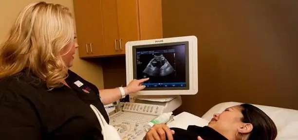
Many women during their first pregnancy are very wary of all kinds of studies, not quite understanding how they go. Screening ultrasound is of particular concern. But you should not worry about this procedure, this is a comprehensive examination that is shown to every pregnant woman without exception. Screening consists of several stages - ultrasound diagnostics and blood sampling from a vein for laboratory tests. The procedure does not require specialized training, but has some features.
Purpose of screening
Screening ultrasound is an absolutely safe, painless and even somewhat pleasant procedure for expectant mothers. Many women treat this study as if they were meeting their child for the first time and are excited about the process ahead.
With the help of screening, the doctor can diagnose the stages of development of the baby and learn about his he alth. Such a study helps to identify pathologies at an early stage. The main purpose of the procedure is to make sure that there are no diseases. If the pathology is still detected, the expectant mother will have to pass moreseveral tests, based on the results of which the doctor will assess the situation and decide whether to extend or terminate the pregnancy.
First screening ultrasound
The duration of the first screening varies from 9 to 13 weeks. This is a very important diagnostic method, which makes it possible to exclude the presence of gross malformations in the fetus. At this time, anatomical characteristics already differ and many organs of the unborn child are visible.

First screening will show
- How many fetuses a woman bears.
- The site of attachment of the placenta.
- The state of the amniotic membranes. If the pregnancy is multiple, then attention is paid to the number of placentas. For twins, they will be separate, and for twins - one common.
- Pregnancy and estimated due date.
- Formation of the umbilical cord.
- Signs of defects on the part of chromosomes. If the fetus has Down syndrome, the screening ultrasound shows an abnormal shape of the nose, a change in blood flow through the tricuspid valve, an increase in the thickness of the collar space.
- Many fetal malformations.
- Pathological conditions, such as signs of placental abruption, threatened miscarriage, etc.
Screening ultrasound in the 1st trimester with the highest quality performance does not give a full guarantee that the fetus has no developmental anomalies. This inaccuracy is due to the too small size of the unborn child. When conducting it, the doctor takes into account such characteristics of the pregnant woman as body weight, chronicdiseases, bad habits. All data is stored in the exchange card of the expectant mother. After completing the study, based on the results, the doctor leading the pregnancy makes a further decision. If there is any doubt, the mother-to-be is referred for ancillary research.
Second screening ultrasound
The second screening allows a clearer assessment of the formation of the fetal organs. The timing of this procedure varies between 19-23 weeks.

Second screening will show:
- Exact gestational age.
- Gender of fetus.
- The presenting part and the position of the unborn child.
- The condition and location of the placenta.
- Amount of amniotic fluid.
- Cervical condition.
At the end of the procedure, the doctor issues a conclusion describing the condition of the fetus. The document also indicates the presence or absence of malformations of the unborn child.
Third ultrasound
The time for the third screening is 32-34 weeks. At this time, they also add caritoco- and dopplerography. These studies provide an opportunity to assess the state of the placenta and the prenatal state of the fetus.

Third screening will show:
- Fetal malformations (if any).
- Previa and position of the fetus. Possible cord entanglement.
- Estimated height and weight of the unborn child.
- Compliance with gestational age and fetal size.
- Structure and functional state of the placenta (suchindicators like thickness, density, maturity).
- Cervical condition.
- Amniotic fluid volume.
- The thickness of the scar on the uterus (for women who have undergone caesarean section).
The result of the screening ultrasound of the third semester affects the determination of the tactics of delivery of a pregnant woman.
Screening results

For the correct interpretation of the screening ultrasound, you need to know the indicators are normal:
- KTP is the size of the fetus from the coccyx to the top of the head. It is indicated in mm. The indicator on the 10th week varies within 33-41 mm, on the 11th - 42-50 mm, on the 12th - 51-60 mm and on the 13th week - 62-73 mm. An overestimated result indicates that the unborn child will be large. Understated indicators are a more alarming sign. Such a result can cause an error in gestational age or indicates the presence of a genetic pathology of the fetus. Also, low results can signal a deficiency of hormones and diseases of the mother.
- TVP - the thickness of the collar space. Its dimensions at the 10th week are 1.5-2.2 mm, at the 11th week - 1.6-2.4 mm, at the 12th week - 1.6-2.5 mm, at the 13th - 1, 7-2, 7. In the presence of genetic pathologies, this figure will be overestimated.
- Nose bone. This indicator can be determined only from the 12-13th week. The result is normal - from 3 mm.
- HR - heart rate. 161-179 bpm at week 10, 153-177 at week 11, 150-174 at week 12, 147-171 at week 13.
- BDP - the distance between the parietal tubercles of the fetus. At week 10, this indicatoris 14 mm, on the 11th - 17 mm, on the 12th - 20 mm, on the 13th - 26 mm. An overestimated indicator indicates a large fetus, but other results should also be above normal. Increased numbers may indicate a fetal brain tumor. Such a vice is incompatible with life. Also, an overestimated figure may be a sign of dropsy of the brain. In this case, by starting timely treatment, you can save the pregnancy.
Whether the result of the screening ultrasound is normal, the doctor determines based on the data obtained after the study.
Preparing for the procedure

For the most accurate test results, a woman needs to prepare for it. First of all, a pregnant woman should tune in to the procedure. To do this, you need to listen to the advice of the doctor leading the pregnancy. He will talk about the features of the procedure and answer your questions. Regardless of the timing of the screening ultrasound, the expectant mother should not be nervous and afraid. The internal state is reflected in the indicators. A pregnant woman should have a few dry wipes or a towel with her in order to remove the remnants of the gel from the abdomen.
Screening consists of two stages (blood sampling from a vein and ultrasound), which are carried out on the same day and in the same diagnostic center (or laboratory).
Before starting the procedure, the pregnant woman is weighed, because some indicators depend on the exact body weight. Also, the expectant mother clarifies all the information about the medications taken,of particular interest are hormonal agents. The day before the ultrasound, a woman is not recommended to have sex, and on the day of the procedure, you can not eat or drink. The moment when it is worth drinking the liquid will be determined by the doctor who will make the diagnosis. Usually a large amount of water is offered to drink an hour before the start of the study. This is done because when the bladder is full, the uterus and, accordingly, the fetus are much better visible.
Is ultrasound dangerous
The first ultrasound was performed over 50 years ago. A lot of experimental and theoretical studies were carried out, which confirmed the safety of diagnostics. This does not pose a threat due to the fact that the baby is protected by the placenta. But if the harm of ultrasound is not proven, then there is no information about its benefits either. Opponents of this research method believe that screening is an unjustified intervention in the female body at the time of the formation of the vital organs of the baby. Also, many doctors do not support ultrasound in order to simply get a photo of the unborn child as a keepsake. Such studies in most cases occur without medical indications.
Officially, ultrasound screening dates start at week 11. It is then that the first procedure takes place, which has not only diagnostic, but also psychological goals. Many pregnant women stop worrying after making sure that everything is fine with the fetus. For many parents, after meeting with the unborn child, new feelings and sensations wake up on the screen of the device. They begin to wait for the baby to appear, realizing thata new role awaits them soon.
When more research is needed

If the expectant mother notices severe abdominal pain, unusual discharge or bleeding, these are signs of a threatened abortion. In such cases, the doctor may prescribe an additional ultrasound.
If a pregnant woman has been ill with any diseases and taken medications, not knowing what the baby is expecting, it is worth doing an additional study. This will give confidence that the drugs did not affect the he alth of the fetus.
Repeated ultrasound is prescribed if it is impossible to determine at the first whether a woman is carrying twins or one child.
An additional study is scheduled at the 13th week if the pregnant woman is diagnosed with complete placenta previa at the 12th week. This is a serious but rather rare complication. In most cases, such a violation occurs in women who have had serious illnesses.
Screening cost
The price of the examination may differ in different regions of the Russian Federation. In private medical centers, the cost for this procedure will be from 1300 to 2800 rubles. A blood test will cost an average of 1,500 to 3,500 rubles.

Women should not forget that pregnancy is not only a wonderful time, but also an alarming condition that requires constant medical supervision. Screening examination as one of the diagnostic methods makes it possible to effectively and timely identify fetal pathologies.
Recommended:
When is the second screening done? Terms, norms, decoding

Examination of a woman's body during pregnancy is mandatory. The performed medical research procedures allow you to monitor the development of the fetus in the womb and, if necessary, correct possible deviations from the norm. This makes it possible to carry a fully developed baby and prevent miscarriage
Norm for screening ultrasound of the 1st trimester. Screening of the 1st trimester: terms, norms for ultrasound, ultrasound interpretation

Why is 1st trimester perinatal screening done? What indicators can be checked by ultrasound in the period of 10-14 weeks?
When do you see twins on an ultrasound? Norms and terms of development, photo

Many women dream of having twins. This is such happiness: your child will never be alone, he will have someone to play with and chat with in the evening before going to bed. Seeing the cherished two strips on the test, many of them run to the doctor, cherishing the hope of hearing the cherished words. And the gynecologist hesitates and waits for something. When do you see twins on an ultrasound? And is everything so clear with multiple pregnancy?
What is BDP on ultrasound during pregnancy: description of the indicator, norm, interpretation of the results of the study

To track all changes and exclude fetal anomalies, its development is monitored using ultrasound. Each time it is necessary to check such basic measurements as BPR, LZR and KTR. What is BDP on ultrasound during pregnancy? Biparietal size - the main indicator that displays the width of the fetal head
Two tests showed two strips: the principle of the pregnancy test, instructions for use, result, ultrasound and consultation with a gynecologist

Planning a pregnancy is a rather difficult process. It requires thorough preparation. In order to determine the success of conception, girls often use specialized tests. They are intended for home express diagnostics of the "interesting situation". Two tests showed two stripes? How can such evidence be interpreted? And what is the correct way to use a pregnancy test? Let's try to figure it all out

