2026 Author: Priscilla Miln | miln@babymagazinclub.com. Last modified: 2025-01-22 17:55:13
The prerogative of modern medicine is early diagnosis. That is why there are scheduled examinations. These include a comprehensive ultrasound of a newborn at 1 month. But why so early? Many young parents may ask this question. This article will help you get the answer to this question.
Examination

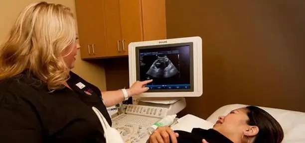
When your baby turns 1 month old, it's time for you to check the baby's he alth. The initial and main study is the diagnosis of the hip joint to detect dysplasia or congenital dislocation. Neurosonography (ultrasound of the brain) and ultrasound of the heart and internal organs (usually the abdominal organs) are also performed. Referrals for these procedures will be given to you by a pediatrician at a children's clinic.
Recently, for reinsurance, many doctors send babies for an ECG (study of heart biopotentials).
In addition to an ultrasound examination, the baby must also be shown to a neurologist, a pediatric surgeon and an orthopedic traumatologist. The rest of the doctors areonly as needed, which is considered in each specific case. But most often a child is also examined by an ophthalmologist, an otolaryngologist and a cardiologist per month.
During the examination, it is advisable to approach narrow specialists with the results so that each of them acquaints you with the norms of ultrasound of a newborn at 1 month.
Importance of the procedure

The first year of a child's life is the most responsible in all development. It is at this time that all the organs and systems of the baby develop and improve. And if this development goes wrong from the very beginning, then it will be much more difficult to fix it later, and in some cases even impossible. The sooner a violation is detected and a correction is started, the more chances there are for a quick relief from a defect or disease without unpleasant consequences.
Therefore, it is in the first year of a baby's life that all vital organs should be examined and unpleasant diagnoses excluded. For this, an ultrasound examination (ultrasound) is performed. It is usually carried out in combination with other tests.
Ultrasound of a newborn at 1 month old allows you to show how the child has adapted to the external conditions of existence and reveal hidden diseases. After all, some anomalies can occur even before the birth of a child, and some in the process of labor.
The prevalence of ultrasound of a baby at the age of 1 month is explained by the fact that this procedure is the safest for such a small person.
Ultrasound of the brain
At 1 month, girls and boys are recommendedundergo a brain exam. It is called neurosonography. It is carried out through the fontanelles - areas of the skull between the bones, covered with connective tissue. They are capable of transmitting ultrasonic waves. Most often, a large fontanel is involved, which is located on the top of the child. Even parents can see it with the naked eye.

All brain structures must be symmetrical, exclude the appearance of neoplasms and changes in the structure. The specialist pays special attention to the cerebral hemispheres and ventricles.
The ventricles are cavities in the brain that communicate with each other and the spinal cord. They contain cerebrospinal fluid that nourishes the brain and protects against damage.
Ultrasound can detect the following diseases at an early stage:
- cysts (fluid areas);
- hydrocephalus (dropsy of the brain, an increase in the amount of cerebrospinal fluid in the ventricles of the brain);
- intracranial hemorrhage;
- ischemic lesions (consequences of hypoxia);
- congenital malformations.
Ultrasound of the heart

Ultrasound of a newborn at 1 month also implies a heart examination. While the baby is in the womb, his heart works a little differently than in an adult. Since the lungs of the fetus are inoperative, it receives oxygen from the mother's blood. This affects the structure and functioning of the child's heart.
BThe structure of the fetal heart has an additional opening, which is called the oval window. A few days after the baby is born, this hole should close up. Ultrasound shows whether this process has occurred. If this does not happen, then this is an indication for registering the child with a cardiologist.
In addition, ultrasound will help to identify other malformations that are not available for detection in other ways.
On ultrasound at 1 month, boys and girls can already reveal some differences in the work of the heart. Girls are known to have faster and more intense heartbeats than boys.
Ultrasound of the hip joints

This examination is performed to rule out hip dysplasia. In this case, the bones that are involved in the formation of the joint are formed abnormally, thereby forming a subluxation or dislocation of the joint.
Most often this pathology occurs in girls (approximately 1-3% of newborns). The pediatrician can already point out the first signs of the disease to you. A child's legs may vary in length, or the folds on the legs may be asymmetrical.
It is in this situation that early diagnosis is crucial. After all, late detection of the disease complicates its treatment and minimizes the chances of a successful recovery.
Various orthopedic appliances, gymnastics, physiotherapy and massage are prescribed as a therapy for dysplasia.
Ultrasound of the kidneys
Does not apply to the number of mandatory examinations in 1 month. When visiting doctors inclinic at the age of one month, the pediatrician prescribes a urine test. If no impurities and pathologies are found, then a kidney examination is not necessary.
However, despite this, kidney disease in newborns is quite common. Approximately 5% of children are at risk. The most common ailment is pyelectasis - an enlargement of the renal pelvis.
If your child has any changes in the functioning of the kidneys, do not be upset ahead of time. Very often, everything normalizes on its own, you just need increased attention to the baby's genitourinary system.
Ultrasound of the abdominal organs
The list of ultrasound of a newborn at 1 month also includes an examination of the OBP (abdominal organs). The liver, pancreas, gall bladder, bladder, kidneys, spleen are examined. All these organs play an important role in the life of a child, so their diagnosis is also necessary.
Where to do an ultrasound of a newborn at 1 month, the pediatrician will tell you. Some even cooperate with private clinics, and therefore they can write you a referral to a specific institution. However, the choice of the place for the examination is still yours, because this is your child.
OBP examination is recommended to be carried out 1.5-3 hours after feeding the baby. Otherwise, the gas in the intestines will interfere with the specialist.
Preparation for examination

Having learned that the child will have a planned examination, parents may be interested in how to prepare for an ultrasoundnewborn at 1 month. Preparation for the examination depends on what kind of ultrasound you are doing.
For example, an ultrasound of the fontanelle, which is included in neurosonography (ultrasound of the brain), is performed without preparation. In addition, there are no contraindications for this either, whatever the condition of the child.
No preparation is needed for ultrasound of the hip joints. Neither the time of feeding, nor the amount of food, nor its ingredients affect the result.
But abdominal ultrasound is performed only after preliminary preparation. To do this, you need to feed the baby and wait 3 hours. That is, it turns out that the examination is carried out on an empty stomach.
If the baby is breastfed, then on the day of the examination, the mother should exclude from her diet those foods that can increase gas formation in the baby (soda, cabbage, legumes).
It is not necessary to clean the intestines artificially (that is, to give the child an enema). This is only permissible when diagnosing children older than 3 years.
Harm ultrasound for baby

Ultrasound of a newborn at 1 month, of course, is a very important and necessary procedure. However, the question arises: "Will the study harm the child?" Parents' concerns are understandable. After all, everyone has heard about the consequences of radiation exposure to the body, so I want to reassure caring parents.
Ultrasound is based on the properties of an ultrasonic wave. There is no penetrating influence of radiation in this procedure. Therefore, there is no harm to the he alth of the baby. That is why this type of diagnostics is used to examine young children from the first minutes of life.
Our grandparents, mothers and fathers say that frequent examinations during pregnancy can harm the baby. It is safe to assure parents, and in particular expectant mothers, that ultrasound during pregnancy can be done without fear for the condition of the fetus. The frequency of ultrasound does not affect the he alth of your unborn child.
Already in the maternity hospital, your child can be examined using ultrasound diagnostics. Since we have found that ultrasound is not harmful, an unlimited number of studies can be performed on a child in one day. On the contrary, for a small person it will be less painful and unpleasant if all the necessary ultrasounds are performed at one time, without stretching it over several sessions.
Recommended:
Stages of vision development in a newborn. Vision in newborns by month

The birth of a child fills your life with a special, completely new meaning. Helpless and tiny, for the first time he opens his huge and slightly surprised eyes and looks into yours, as if saying: “You are my whole world!”. The very first smile, the language of communication that only the two of you understand, the first word, steps - all this will be a little later. The basis of future achievements is the correct formation of all systems and organs. In this article, we will take a closer look at the stages of vision development in a newborn
Norm for screening ultrasound of the 1st trimester. Screening of the 1st trimester: terms, norms for ultrasound, ultrasound interpretation

Why is 1st trimester perinatal screening done? What indicators can be checked by ultrasound in the period of 10-14 weeks?
2-month-old baby: daily routine. Development of a 2 month old baby

Here is your 2-month-old baby who has changed so much in such a short period of time that you no longer know what will happen next. From this article you will learn how to care for your little one, how the baby should develop properly, what daily routine suits him best
How to prepare for pregnancy? Do I need to prepare for pregnancy?

Pregnancy is a very important period in a woman's life. At this time, the formation of the unborn child takes place. Its development directly depends on the lifestyle of the expectant mother, as well as on the required amount of vitamins and minerals. Their lack can provoke the occurrence of various defects and malformations. In this regard, it is extremely important to take a responsible approach to the stage of pregnancy planning
How to prepare for the wedding and where to start? Stages by month

A wedding is a very exciting event that will forever remain in the memory of not only the newlyweds and their parents, but also the guests. In order for the day of its holding to be remembered only by pleasant and bright moments, it is necessary to prepare in advance for it. How to prepare for a wedding? Where do you need to start and what elements that make up the celebration should you pay attention to? More on this later

