2026 Author: Priscilla Miln | [email protected]. Last modified: 2025-01-22 17:55:29
How to find out if the fetus is developing correctly, are there any deviations, how are the internal organs of the crumbs formed? The answers can be given (when the period to which your pregnancy has come - 12 weeks) ultrasound. Screening allows you to assess the development of the fetus, gives a clear picture of the genetic and chromosomal characteristics of the future baby. This makes it possible to determine the presence or absence of anomalies.
Ultrasound at 12 weeks

Basically, the procedure is carried out in two ways: transvaginally (through the vagina using a special sensor) and transabdominally (through the skin of the abdomen). The latter is more common, and the former is not prescribed to all women in position, but only to some of them, in cases:
- if the placenta (or chorion) is low attached;
- if there is isthmic-cervical insufficiency, and it is necessary to assess its degree;
- if there are signs of inflammation of cysts and appendages (to accurately establish the diagnosis), or the nodes of uterine fibroids are very specifically located, and method No. 2 showed little information;
- when assessing the collar zonebaby or measurements of the right size that are difficult to take due to the fact that the fetus is not positioned as it should be, or the subcutaneous tissue of the abdomen is very thick.
The study is being conducted in this way: a woman lies with her knees bent; The doctor inserts an ultrasonic transducer into the vagina and covers it with a disposable condom for protection. Usually everything is done with great care, so the pregnant woman does not feel pain.
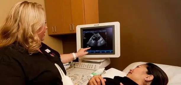
Transabdominal examination done in the same position. All the air between the transducer and the skin will not be expelled, so incorrect results may occur. To reduce the chance of error as much as possible, a special gel is used, which is applied to the abdomen. Gradually move the sensor across the abdomen so that you can see the organs of the crumbs, as well as the mother's uterus and placenta. Ultrasound is completely safe for the fetus and does not cause any damage to it.
How to prepare for an ultrasound
Preparation depends on the method. If transvaginal is used, then it is recommended not to consume 1 day before the study those foods that can cause fermentation: white bread, legumes, cabbage, peas. The intestines must be emptied, otherwise the gases present there will interfere with the examination of the uterus and fetus. If there is a feeling that the stomach is swollen, you can drink the drug "Espumizan", which is harmless to the fetus.
Before a transabdominal examination, drink half a liter of water 30 minutes before the start. This is necessary to have a full bladder, which will allow you to examine the fetus and assess its condition.

Child development on12-week period
Many of the main organs of the baby have already developed, and some small structures continue to form. On average, a child is 80 mm tall and weighs about 20 grams. Doctors also note that the fetus has the following features:
- heart rate is more rapid than in the third trimester, and can be approximately 170 beats per minute;
- the baby's face no longer looks like a tadpole, but takes on human features;
- you can see the eyelids, lobes, a little fluffy hair (at the site of the formation of eyebrows and eyelashes);
- most of the muscles have practically already developed, so the fetus moves all the time, and the movements are mostly involuntary and rather chaotic;
- baby grimaces and clenches her hands into fists, nails can be seen on her fingers;
- the child has already developed kidneys and the intestines are almost formed, red and white blood cells are observed in the blood;
- both hemispheres of the brain are fully formed, however, while the spinal “commands”;
- you can see who it is: a boy or a girl, but since the fetus does not always lie as the mother and doctors want, you can make a mistake, so they say more precisely about the sex at the 16th week.

How to read the results?
You will receive papers with the results of the study after the screening is done (12 weeks). A transcript of the analysis will be given below.
Starting from the third month, it is already clearly visible whether one child or not. Therefore, if in the column“number of fetuses” is written two or more, this indicates that you will have twins (triplets, etc.) You can also already find out if the fetuses are identical (twins) or twins (heterozygous).
Previa
This is the name of the part of the fetus closest to the birth canal. At 12 weeks, it can be anything: legs, head, or the baby is completely diagonal. The final presentation is assessed at the 32nd week of pregnancy. If the head is not located towards the exit from the uterus, then all possible measures are taken to correct this situation.
Measuring the size of the fetus (or fetometry)
Deciphering the ultrasound is needed to evaluate the parameters, but this should be done by a doctor who will be guided not only by the numbers, but also by the general situation of the pregnant woman. All norms are designated by certain letters and numbers. Here are the main ones:
- BPR (BPD, BRGP) - this abbreviation denotes the so-called biparietal size, i.e. the distance of the head from one parietal bone. At 12 weeks, the ultrasound should show 21 mm BDP.
- Baby's height is approximately 8.2 cm, weight should not be less than 17-19g.
- FML, DLB is the length of the thigh. The norm is from 7 to 9 mm.
- The collar space should not exceed 2.7 mm. By its size, it is determined whether there are any serious illnesses. On average, it is approximately 1.6 mm.
- The term KTP (CRL) denotes the coccyx-parietal size, i.e. the maximum length from the head to the tailbone, the norm is 43-73 mm.
There are also other abbreviations:
- HUM (DP) -shoulder length.
- AC (OJ) - abdominal circumference.
- ABD (J) - belly diameter.
- RS - heart size.
- OD - head circumference.

For all these parameters, 1 screening during pregnancy allows the sonologist to determine how the baby's structures grow and develop. If the measurements made are less than the norm, then according to the total population, they evaluate how they decreased: proportionally and simultaneously or not. If they do not coincide only slightly, then there is no reason to panic. Perhaps the deadline was incorrectly determined, and in fact it is only the 11th week. Or perhaps the baby is so tall because of short parents.
They also find out if there are any malformations in the development of internal organs, is there an entanglement of the umbilical cord, what is the heart rate (the norm is from 150 to 174 beats per minute), are there any deviations in the characteristics of amniotic fluid.
Reading the conclusion of an ultrasound scan, a pregnant woman may encounter such concepts as "polyhydramnios" and "oligohydramnios". What is it and is it something to be afraid of? There is nothing wrong with these words. This is just a determination of the amount of those waters in which the fetus swims: if there are more of them than necessary, polyhydramnios is fixed, if less - oligohydramnios. Often this indicates some kind of violations: intrauterine infection (IUI), impaired functioning of the kidneys, the central nervous system. Also check if the water is cloudy. If so, this is a clear indication of an infection.
The basic rule when detecting deviations from the norm is not to panic, but to go tospecialist.
Can there be deviations from the placenta?
Ultrasound shows where the "baby place" is attached, how mature it is, whether there are pathologies and more. The best option is to attach to the back wall of the uterus. But the placenta can "cling" to the front, and even to the bottom. However, it should not overlap the internal os of the uterus. This condition is called chorionic, or central placenta previa. In this case, they monitor whether the situation will change, and if not, then a caesarean section is performed for delivery. If the pharynx is not completely covered, it is called an incomplete presentation; childbirth is carried out in the usual manner.
If the placenta "settled" near the exit (less than 70 mm), then this is a low presentation. Since it can become a threat of bleeding, a less active regimen is recommended for a pregnant woman. Then they observe whether the placenta rises up. If this happens by 32-36 weeks, then there will be no threat, and the woman will give birth in the usual way.
The maturity of the placenta at this time is 0. The "lobular" placenta is the second degree of maturity, and in such a situation, you should consult a doctor. Deposits of calcium s alts are called calcifications. It is considered normal if they are present in the placenta of the first degree of maturity.
If there is a death of some part of the "children's place", this is called a placental infarction. In this case, you urgently need to consult a doctor to find out the cause and prescribe treatment, because if this continues to happen, the child will not have enough oxygen and the necessarydevelopment of substances.

Cervix: condition, structure
At the 12th week, the size of the cervix is measured, which should not be shorter than 30 mm. The longer it is, the better. If it is very short, less than 20 mm, then the pregnant woman is hospitalized, and possibly surgery will be used for treatment. The os of the uterus must be closed, both external and internal.
Myometrium (or muscle condition) shows if there is a risk of miscarriage. If the diagnosis indicates that at this time there is uterine hypertonicity, then the woman is treated. Especially alarming are such facts as the "petrification" of the abdomen, "push-pull" in the lumbar region.
How is the term determined by ultrasound
Using special tables, the KTR calculates the gestational age. It may be that such a function is built into the program of the ultrasound machine. Compare the terms - calculated from the last menstruation and issued by ultrasound. If the difference is small (one or two weeks), then the exact period determined by the obstetrician is considered. In case of a greater discrepancy (more than 2 weeks), the period determined by ultrasound is taken as a given.
Prenatal screening: what it is and how it is done
You should be especially careful when the pregnancy is 12 weeks. Ultrasound, screening - all these studies are designed to assess the development of the fetus. At the same time, ultrasound is done first, and then screening is already prescribed (depending on the indicators). Spend it if:
- Pregnant 35 years or older.
- Before this, dead babies were born.
- When examining previous fetuses, intrauterineinfection.
- A baby was born with a chromosomal abnormality.
- It has been established that relatives of both parents have such anomalies.
Only special centers screening (12 weeks). How do they do it? They collect all the tests: ultrasound, blood, external data. The evaluation of the study is done by a geneticist, and attention is mainly paid to the collar and these indicators: free β-hCG and PAPP-A. Basically, these markers are studied in a well-established combination. If at least one of them has changed, this does not mean at all that the fetus has some kind of pathology.
So when screening is done at 12 weeks pregnant, the characteristics of these markers are used. These are whey proteins. If they have deviations, then the child will have genetic disorders. Free β-hCG is a subunit of human chorionic (chorion is a germ) human gonadotropin, and PAPP-A is a pregnancy-associated protein A. To study these indicators, ELISA (enzymatic immunoassay) analysis is used.
HCHG stimulates the synthesis of steroid hormones (in the placenta and corpus luteum). Doctors have already found out that it is hCG that protects the fetus from rejection. By examining its level, one can make predictions for the further course of pregnancy. According to medical statistics, hCG gradually increases until the 10th week, and then remains at about the same level (from 5000 to 50000 IU / L) until the 33rd week, after which it may rise slightly.
1 Pregnancy screening is done between the 10th and 13th weeks of your due date. To calculate all the risks, they take a lot of data: the date of ultrasound, KTR and TPV (collar thicknessspace).

These analyzes are very important for determining the existing pathologies in the chromosomes. However, if the readings are slightly increased, do not worry and draw hasty conclusions. You just need to turn to a geneticist who will tell you what to do next. There is also a possibility that the ultrasound was misread. Screening for a 12-week pregnancy can be repeated - for clarification, or the doctor will prescribe an invasive diagnosis that will more accurately determine the genetic makeup of the child. Depending on how long it takes, either a chorionic villus biopsy or amniocentesis is done.
If even 1 screening showed a very low risk of chromosomal pathologies in the fetus, then you should not refuse the examination conducted at 4-5 months of pregnancy. In addition to hCG and AFP, the level of free estriol is determined (triple test).
In order to determine the indicators of β-hCG and PAPP-A, donate blood for screening. 12 weeks is already a sufficient period for biochemical analysis to reveal the presence (or absence) of abnormalities in the chromosomes.
Conclusion on analyzes
Depending on the results of the blood test, it is revealed why the indicators differ from the norm. For example, a 12-week pregnancy screening may reveal the following:
- Down Syndrome.
- Not one fruit, but 2 (3, etc.). More fruits - more hormone levels.
- Toxicosis.
- Wrong gestational age. For each week of development of the child corresponds to some indicator,determining the exact age of the fetus.
- The presence of diabetes in the mother.
- Ectopic pregnancy.
- High risk of miscarriage.
Are there any performance standards?
Of course there is! You can find out by doing such studies as ultrasound, screening (12 weeks). The norm will become known after studying the data by the doctor. However, there are average medical indicators clearly established for each week of pregnancy. For example, β-hCG at 11-12 weeks should be between 200,000 and 90,000 mU/ml.
However, it should be borne in mind that screening for a 12-week pregnancy gives, of course, very high, but still not one hundred percent results, because each woman has her own body characteristics, which are necessarily taken into account by the doctor. If the fetus is not one, then it is more difficult to diagnose. Look at indicators. If they are one and a half or two times larger, then we can conclude that there are 2 or more embryos. Each of the fruits has its own chorion and different hormone production. Therefore, the figures are so high, and the expectant mother is sent for an ultrasound scan to confirm a multiple pregnancy.
As soon as the screening is done (12 weeks), the normative values are immediately checked against the data obtained to calculate if there are any pathologies. Physicians for these purposes use a special coefficient called MoM. It is calculated according to a certain formula: the amount of the hormone that was determined by the results of the screening is divided by hCG (corresponds to the norm during this period of pregnancy). You should get a unit (this is ideally). Wellaccording to the results of all studies, it is judged whether to include the expectant mother in the risk group with chromosomal abnormalities or not. It is worth noting that even if this suddenly happened, this is not the final verdict, but only one of the probabilities. Therefore, the rest of the indicators are compared and only after that do some conclusions. Reviewing all 12-week screening: Ultrasound, hormones, TVP, may re-test in 2nd trimester.
PAPP-A protein is responsible for the immunity of a pregnant woman, and it also helps the placenta to work. Since the boundaries of the thresholds are clearly established, its deviations are highly undesirable. The thing is that such "jumps" of indicators speak not only of a possible miscarriage, but also of such terrible anomalies as Down's syndrome, de Lange's syndrome, etc. Such numbers are considered normal: from the 11th to the 12th week - 0.7-4.76; from the 12th to the 13th week - 1, 03- 6, 01.

Research Feedback
Women who have had screening (12 weeks) have varying opinions about it. Some have misidentified the gender of their baby. There is an explanation for this - the period is too short, it will finally be possible to say who will be born: a girl or a boy, only at the 16th week. They also talk about different prices. Some take tests for free, others pay from 1,000 to 3,000 rubles.
However, most mothers note that ultrasound and screening help to understand how the baby is developing. Since now these procedures are mandatory, it is possible to diagnose and start treating existing diseases in time tothe baby was born he althy.
Recommended:
How to behave during the first weeks of pregnancy. What not to do in the first weeks of pregnancy

In the early stages of pregnancy, you need to pay a lot of attention to he alth. During the first weeks, the tone for the subsequent course of pregnancy is set, therefore, the expectant mother should especially carefully listen to her feelings and take care of herself
Norm for screening ultrasound of the 1st trimester. Screening of the 1st trimester: terms, norms for ultrasound, ultrasound interpretation

Why is 1st trimester perinatal screening done? What indicators can be checked by ultrasound in the period of 10-14 weeks?
Discharge at 30 weeks pregnant - what to do? 30 weeks - what's going on?

Here comes the 30th week, 2/3 of your pregnancy is already behind, and ahead of childbirth, meeting with the baby and many positive moments. To warn yourself against negative aspects (such as pathological discharge at the 30th week of pregnancy and, as a result, premature birth) or at least minimize them, you must follow the basic rules and tips
Delivery at 37 weeks of gestation: the opinion of doctors. How to induce labor at 37 weeks?

Pregnancy is a very responsible period for every woman. At this time, the body of your crumbs is formed and develops. In many ways, the he alth of the future depends on the course of pregnancy
The norm of hCG during pregnancy: table and transcript

Nowadays, it is not difficult to establish the fact of pregnancy, since pharmacies sell funds specially designed for this. We are talking about tests that are in different price categories from the cheapest to the most expensive. But if the results are not satisfactory, and the ultrasound cannot give a definite answer, then you can donate blood for analysis, where the hCG rate will be determined. Moreover, this hormone is found not only in the blood, but also in the urine of pregnant women

