2026 Author: Priscilla Miln | miln@babymagazinclub.com. Last modified: 2025-01-22 17:55:13
For most people around, pregnancy becomes obvious only from the moment when the mother's tummy noticeably rounds. However, already from the first week after conception, changes imperceptible to the eye begin to occur in the female body. During the examination, the gynecologist can determine the onset of pregnancy by the increased size and location of the uterus. By weeks of pregnancy, an accurate description is provided only according to the results of an ultrasound examination. Now it is assigned to every woman who has a baby.
Consider the location of the uterus by week of pregnancy. The photos presented in the article will help to explain this clearly. Thanks to many years of experience, it was possible to determine the norms and criteria for assessing the size of the main reproductive organ (uterus) from conception to childbirth. Consider these figures.

Sizes at different dates
Self-determinethe location of the uterus by week of pregnancy in the first trimester is quite difficult. Moreover, you should not do this, so as not to provoke the tone of the reproductive organ, which can lead to miscarriage. As a result of palpation, the doctor can determine that the body of the uterus is enlarged. If we talk about the first month of pregnancy, then the reproductive organ resembles a chicken egg in size, and after a month it becomes like a goose in terms of parameters. The size of the fetal egg for a period of 10 weeks is 22 mm, and the volume of amniotic fluid is 30 ml.
Every day the growth of the reproductive organ becomes more and more obvious. Your doctor may need a tape measure to determine the location of your uterus by week of pregnancy. The photo below shows how the indicators change. The tape will allow during the next examination to obtain information as to whether the growth of the fetus corresponds to the obstetric period. Such a measurement also allows you to establish the presence of oligohydramnios or polyhydramnios.

Shape of the uterus
The reproductive organ changes depending on the gestational age. For the first two months, its shape resembles a pear. The embryo has enough space inside the ovum, since its size is still extremely small to affect the appearance of the uterus. But by the end of the third month, it becomes more spherical. Its shape becomes even more ovoid as the placenta develops. She remains like this until the birth itself.
Bottom height

To the doctorcould determine the location of the uterus by weeks of pregnancy, he is guided by an ordinary centimeter tape. Determination of the standing height of the fundus of the uterus (VDM) is necessary in order to be able to establish without an ultrasound examination whether the pregnancy corresponds to its real term. According to obstetric practice, this indicator helps to determine how many centimeters the bottom of the uterus rises at each gestational age. The measurement begins in the second trimester, when the reproductive organ begins to protrude noticeably. For example, if you take the location of the uterus at the 15th week of pregnancy, then the doctor on the centimeter tape should fix the VMD, which is 15 centimeters.
How the uterus grows in the first trimester
The first months of pregnancy for many women go unnoticed. Therefore, they do not even know what changes are taking place in the uterus. When planning a pregnancy, a woman can follow how the location of the reproductive organ changes with the help of ultrasound.
It is advisable to exclude frequent visits to an ultrasound specialist, as this is associated with a certain discomfort for the expectant mother. However, in some cases this measure is forced. In particular, already at the 6th week of pregnancy, its further development can be determined by the location in the uterus of the embryo. If it is attached to the lower wall, then there is a risk of complications or a threat to gestation. The expectant mother should be more careful and avoid physical exertion. By the end of the third month (first trimester), the formation of the placenta begins.
Secondtrimester
At the 16th week of pregnancy, the location of the uterus can be roughly determined between the navel and the pubic bone. After 4 weeks, WMD is two fingers below the navel, and a noticeably rounded tummy is already difficult to hide from others. The size of the fetus during this period is about 26 cm, and the weight is 270-350 g. At this time, the child is quite comfortable inside, so the mother can feel his physical activity during the day.
Therefore, the location of the baby can be head, pelvic or transverse. The latter affects the location of the uterus, but is not a cause for concern. In order for the baby to return to its usual position in the uterus, at the 18th week of pregnancy, walking in the fresh air, as well as performing special exercises, can be recommended. The baby can return to the correct position on its own if the mother helps him with this. Doctors recommend getting on all fours. This makes it easier for the baby to roll over.
At the 20th week of pregnancy, the position of the uterus inside the abdominal cavity becomes even more dominant. Mom begins to feel more obvious movements.
In the second and subsequent pregnancies, doctors and many mothers note the first tremors at an earlier date. At 24 weeks, the bottom of the uterus reaches the navel, a month later it rises two fingers above it. At this time, the baby is intensively gaining in height and weight, taking up more and more space inside the mother's womb. Consequently, there are significant changes in the size of the uterus itself. It stretches more and more, and during the movements of the baby, the mother can even feelhow it touches the internal organs.
Amount of amniotic fluid and uterus size

At 12 weeks of pregnancy, the doctor can determine the location of the uterus by palpation, it is felt through the abdominal wall. Its dimensions at this time are comparable to the head of a newborn baby, and the VDM is located in the region of the upper edge of the pubic bone. Closer to 16 weeks of pregnancy, the formation of the placenta ends, which protects the baby and is a filter for determining his he alth status.
In the first trimester, the amount of amniotic fluid is very small. However, by the end of the second trimester, this figure is approximately 600 ml. By the third trimester, the volume of water is about 1.5 liters. Knowing the norms, we can talk about a lack or excess of amniotic fluid.
If a woman notes that her belly is smaller or larger than expected, then it may be polyhydramnios or oligohydramnios. However, do not self-diagnose. During the examination, the doctor can only suggest a particular pathology. An accurate result can be obtained during an ultrasound examination.
Third trimester
By 32 weeks, the fetus is about 42 cm tall. Consequently, the mother's tummy at this time is already quite impressive. Its circumference is about 80-85 cm, the navel is smoothed out. The location of the fetus at this time in most cases is head down, but even with the pelvic position, it is likely that the baby willflip over.
This period is the time for the third screening, which includes another ultrasound. It allows the doctor to determine the week of pregnancy by the location of the uterus, to find out changes in the parameters of the child, to clarify whether his weight and height correspond to the term. The height of the fundus of the uterus should be within 31-33 cm, and at 34-35 weeks - 32-33 cm.
Parameters in the last month of pregnancy

After 36 weeks, labor can begin at any time. In some cases, women note that the stomach drops down. This is due to the fact that the baby changes his position, moves less (he is already large, so he is cramped in the womb) and prepares for the birth. By the end of the eighth month of pregnancy at 38-39 weeks, the WDM norms are 35-38 cm.
If at 41 weeks of pregnancy the position of the baby in the uterus is wrong, then there is practically no chance that he will turn his head down. Therefore, doctors usually recommend a caesarean section.
The height of the fundus of the uterus at this time becomes somewhat less - about 34-35 cm. This indicates that the baby is preparing to be released, and labor can begin at any time.
Uterine size in multiple pregnancies
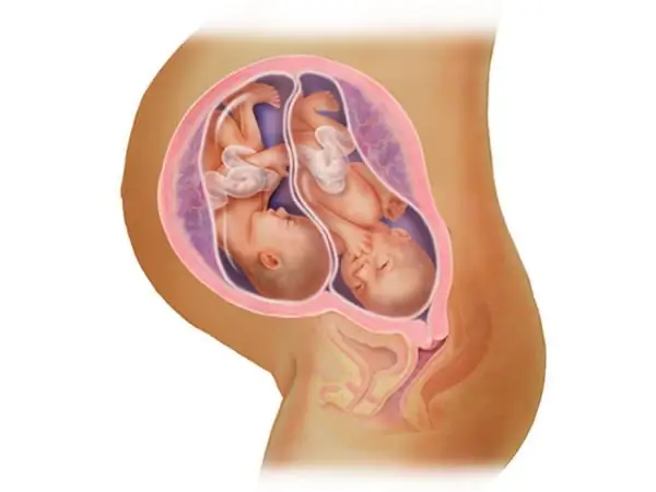
For expectant mothers who monitor changes in their body, doctors recommend not to panic if the location of the uterus at 10 weeks of pregnancy is slightly larger than others. Perhaps they are expecting twins or triplets. Dimensionsthe uterus and abdomen in multiple pregnancies are noticeably larger. The growth of each of the babies is about 10 cm. You can confirm or refute this fact during a routine screening. This is especially true for those who did not have an ultrasound before the end of the first trimester.
Ultrasound to determine the location of the fetus in multiple pregnancies

Since the load on the body is growing with a double force, you should be prepared for the fact that pulling pains in the lumbar region will often be felt. The sprain that occurs in connection with the growth and change in the position of the uterus also cannot pass without a trace. By the 18th week of pregnancy, doctors and experienced mothers recommend wearing a bandage. This will reduce the likelihood of improper positioning of babies inside the womb and distribute the load, which is caused by a shift in the center of gravity. By week 20, the weight of each baby reaches 400 g, and after two weeks (if the pregnant woman is carrying twins), the total weight is more than 1 kg.
Women who have multiple pregnancies, it is not easy to determine the exact period without a special study, since their belly size is somewhat larger than that of those who bear one baby. Ultrasound allows you to determine the correct location of the uterus by weeks of pregnancy, which is very important for the doctor, because it makes it possible to assess how the babies feel inside the mother's womb. The same diagnostic method allows you to identify the presence of abnormalities or developmental delays, which occurs when carrying twinsoften.
Mid Pregnancy
In the second trimester, you can listen to the heartbeat of babies. This will help to establish their location without the use of ultrasound. The doctor uses a conventional phonendoscope (a tube made of wood or metal) or a familiar stethoscope. Such a procedure for a singleton pregnancy is resorted to from the 20th week.
However, for moms with two or more babies, this period can be shifted a few weeks earlier. Judging by the reviews of experienced mothers, the tummy begins to grow from the 12th week, and the first movements are felt at the 17th week of pregnancy. The location of the uterus at this stage is somewhat different than in a singleton pregnancy, because having multiple babies requires more space inside the reproductive organ, so it has to stretch faster.
At the 26th week of pregnancy, an enlarged uterus can cause significant discomfort. Long walks quickly tire the expectant mother. The weight of her children is about one and a half kilograms.
Difficulties in determining the location of the fetus

By the end of the second trimester, the doctor can feel the location of both babies inside the womb through the stomach. However, there are also difficulties here. For example, if the second baby is hiding behind the first (closer to the spine), it is very difficult to listen to his heartbeat and determine the location.
In this case, there is only one way - ultrasound, which accurately shows their location in the uterus by week of pregnancy.
Last trimester multiple pregnancy
In the third trimester, special attention is paid to what position the kids have taken. This affects the further choice of the method of delivery. The height of the fundus of the uterus at 8 and 9 months may not differ much from each other. There is very little room for movement in the reproductive organ. It often happens that one fetus is in the correct position (head down), and the other is incorrect. When listening to the heartbeat, the doctor can easily determine the location of the children. However, an ultrasound is prescribed to be sure.
In medical practice, multiple pregnancies end at 39-40 weeks. The size of the abdomen becomes quite impressive, it is difficult for a woman to move around. As a rule, after 36 weeks, a decision is made to perform a caesarean section.
Recommended:
Development, weight and size of the fetus at the 16th week of pregnancy

Development, weight and size of the fetus at the 16th week of pregnancy. What happens during this period with the child and the expectant mother? How does her state and feelings change? General tips and tricks for girls in the 16th week of pregnancy
Changes in the uterus during pregnancy

What happens to the uterus during childbearing? Anatomy, functions, features of growth and work of the most important organ of the female reproductive system during pregnancy
Feelings at the 7th week of pregnancy: norms of fetal development, woman's feelings and changes in the body

After pregnancy is confirmed, the woman is aware of her new position. She listens to all the sensations, wonders if the fetus is developing normally. At each stage of pregnancy, certain symptoms occur. They can tell about the state of the woman's body. What sensations at the 7th week of pregnancy are considered normal, what happens to the body of the expectant mother and the fetus, will be discussed in the article
9 week of pregnancy: weight and size of the fetus

From 7 to 9 weeks of pregnancy, there are many processes and transformations in the womb of the expectant mother. And although a clear increase in the abdomen has not yet been observed, the development of the fetus is carried out at a fairly rapid pace: the weight is 7 grams, the height is 4 centimeters. And by the 9th week, he already has all the makings of a future person (in terms of the body, systems and organs), only in miniature. All this is a real miracle that the expectant mother comes into contact with. For more information on this topic, see our article
Second week of pregnancy: signs and sensations, fetal development, abdominal circumference and changes in a woman's body

Pregnancy from its first days until childbirth is a bright and wonderful process. Many mothers become interested in what is happening with their body, because a global restructuring begins, what changes are observed, sensations. It is worth having a clear idea of what the normal state is and what you should not be afraid of at first, because in case of any deviations, you should consult a doctor

