2026 Author: Priscilla Miln | miln@babymagazinclub.com. Last modified: 2025-01-22 17:55:13
It would seem that studies in the early stages of pregnancy, which determine and prevent the development of pathologies, are of particular importance. But ultrasound in the last stages reveals a number of factors that indicate the readiness of the fetus for childbirth. One of them is the Beklar core.
The importance of late-term ultrasound
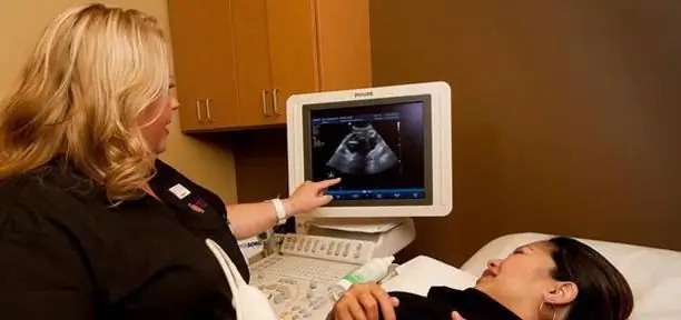
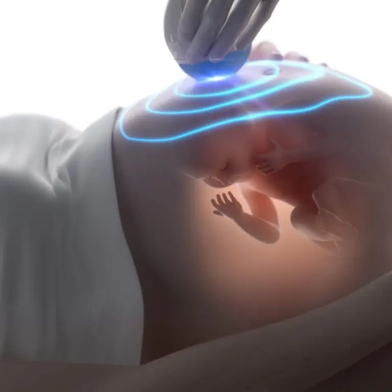
Elective ultrasound in the third trimester is scheduled for 31-32 weeks, but in some cases, doctors may prescribe a prenatal examination. For example, against the background of problems (pulling pains in the lower abdomen, stained with an admixture of discharge blood). Or if the expectant mother is at risk (age, he alth problems). Based on the ultrasound results, the attending physician will draw certain conclusions:
- Birth presentation - the position of the baby in the uterus will tell you the strategy for conducting the birth process.
- Readiness of the placenta for childbirth - depending on the maturity and thickness of the placental membrane, more accurate terms of the PDR are set.
- Diagnosisdevelopment of the respiratory system will indicate the degree of term of the child.
- Pathologies - it is assessed if there are any pathological changes that threaten the condition of the fetus.
- Anatomical parameters - examine the heartbeat, frequency of movements, the maturity of the liver and other internal organs, the height and weight of the fetus, the presence of ossification nuclei.
As you can see, the control of these indicators is important for the successful completion of childbirth and the he alth of the baby and mother.
What does the Beklar core show during pregnancy
In ultrasound examinations performed at 37-40 weeks, certain parameters deserve special attention. One of the anatomical indicators that specialists keep under control is the Beklar nucleus, which is the degree of ossification of the distal epiphysis of the femur. According to medical encyclopedias, it is an important sign of full term.
Often the concept of Beklar's nucleus is confused with the ossification nuclei in the hip joint. The formation of these nuclei begins earlier - in the range from the third to the fifth month of pregnancy, during the period of active osteogenesis. Difference in localization: Beklar's nucleus occurs in the lower part of the thigh bones.

Normal values at 37-40 weeks
The values of the nucleus of ossification of the distal epiphysis in the fetus vary from 3 to 6 mm by the fortieth week of pregnancy. These dimensions are considered a sign of the norm. However, in 3-10% of cases of normal full-term newborns, Beklar nuclei were absent altogether, and in some cases their formation was observed already at 35-36week.
Thus, it is wrong to judge the maturity of the fetus only by the size of the Beklar kernel. The decision on the presence of pathology is made by the doctor based on deviations in the totality of parameters.
Measures taken in case of abnormalities
Normally, Beklar's nucleus completes its formation after the birth of a child, by the sixth month of life. What threatens insufficient ossification?
Firstly, the delay in ossification of the lower epiphysis of the femur entails the pathology of the development of the knee joint. As a result, the baby cannot crawl normally.
Secondly, there is a risk of developing epiphyseal dysplasia. At the same time, a large role of molecular genetic disorders is noted in the pathogenesis.

Distal epiphyseal dysplasia causes X- and O-type leg curvature. Deformities, thickening or expansion of the knee joints occur. Short stature is also noted, caused by a reduction in the length of tubular bones. According to experts, these problems do not affect the life expectancy or intellectual development of the child. However, they can cause hypoplasia of the vertebral bodies and shortening of the spine. At the same time, ossification of the vertebrae is delayed.
Diagnostics are carried out with the help of X-ray studies, molecular genetic tests and regular visual examination. Deceleration of ossification in the lower epiphyses is detected primarily on radiographs.
Treatment of dysplasia in the early stages of a child's development is limited to supportive and corrective therapy. It is important to use orthopedic devices: bandages and corsets reduce the load on the spine and joints. For babies, therapeutic massage and exercise therapy are effective.

Surgical correction of deformities is possible in adulthood.
Timely identification of the problem and the presence of orthopedic treatment help to reduce the negative manifestations of the disease.
Recommended:
Can an ultrasound not show pregnancy? Fetal size by week of pregnancy

There are times when women find out they're pregnant when they're well into their term. There are quite a few ways to confirm a special situation using the analysis of hCG, various tests. But sometimes, the listed methods do not always carry reliable information. Can an ultrasound not show pregnancy? We'll talk about it in this article
Teenager is a special system of values

Teenager is not just a word for a teenager of 13-19 years old, it is a whole culture and system of life values, inextricably linked with certain problems and social phobias
Norm for screening ultrasound of the 1st trimester. Screening of the 1st trimester: terms, norms for ultrasound, ultrasound interpretation

Why is 1st trimester perinatal screening done? What indicators can be checked by ultrasound in the period of 10-14 weeks?
Lymphocytes in children are normal. Lymphocytes in children (normal) - table

A blood test is prescribed to make sure the presence or absence of various diseases. There are white and red cells in the blood. Lymphocytes are white cells. Experts pay special attention to their number, as they can indicate very dangerous diseases. How many should there be and what is the norm for children?
Should I do an ultrasound in early pregnancy? Pregnancy on ultrasound in early pregnancy (photo)

Ultrasound came into medicine about 50 years ago. Then this method was used only in exceptional cases. Now, ultrasound machines are in every medical institution. They are used to diagnose the patient's condition, to exclude incorrect diagnoses. Gynecologists also send the patient for ultrasound in early pregnancy

