2026 Author: Priscilla Miln | miln@babymagazinclub.com. Last modified: 2025-01-22 17:55:29
Ultrasound came into medicine about 50 years ago. Then this method was used only in exceptional cases. Now, ultrasound machines are in every medical institution. They are used to diagnose the patient's condition, to exclude incorrect diagnoses. Gynecologists also send the patient for an ultrasound scan in the early stages of pregnancy. However, women are skeptical of such studies. The opinions of doctors on this topic also differ. Let's try to figure out whether it is possible to do an ultrasound in the early stages. You will also find out who really needs to undergo the indicated diagnostics.

Types of diagnostics
Before you go for an early ultrasound, there are a few things you need to know about this type of examination. Diagnosis can be carried out by two methods - vaginal and through the abdominal wall. At the same time, specialists use different sensors. ATIn public institutions, research is usually free of charge. A woman should have only a passport and an insurance policy. If you go to a private clinic for diagnostics, you will have to pay for the procedure. The average cost of manipulation is from 500 to 2000 rubles, a lot depends on where you live, the qualifications of the doctor and the modernity of the equipment.
It is worth noting that the use of vaginal research methods on ultrasound in the early stages allows you to get more accurate information. In the first weeks after conception, manipulation through the abdominal wall may not show any important details at all. The doctor should choose the diagnostic method. Sometimes both methods are used. In this case, the specialist receives maximum information.
Preparation for the procedure: general description
How is an ultrasound done in early pregnancy? For diagnosis through the abdominal wall, a woman is asked to drink two glasses of water a few minutes before the procedure. When filling the bladder, the reproductive organ is better visible. For examination, you will need to expose the lower abdomen.
If you have a vaginal ultrasound, you should definitely take a tissue with you. It is also worth taking hygiene measures before the examination. Remember the date of your last menstruation, or rather, the day it began. The doctor will definitely ask you for this information. Each clinic may have its own additional conditions for the study.

Should I do an ultrasound in the early stagespregnancy?
This question remains controversial to this day. The opinions of doctors are rather ambiguous. It all depends on each specific situation. Some women are not advised to conduct diagnostics until 12-14 weeks. It is during this period that a planned study is carried out. Other expectant mothers are strongly recommended to visit the ultrasound diagnostic room.
Women's opinions also differ on this issue. Some representatives of the weaker sex are sent for research immediately after a delay in menstruation. Other persons do not want to conduct an examination even by the end of the first trimester. In any case, if you do not go for an ultrasound in the early stages, then you should definitely make a diagnosis according to the screening time.

Examination time
When to go for an ultrasound during early pregnancy? If you go to the doctor immediately after the first day of the delay, then the diagnosis will not show any result yet. Even the most modern equipment is unable to fix a fetal egg less than one millimeter in size.
To establish the fact of conception, you need to visit the ultrasound room about one week after the delay. If you want to make sure that the baby's heart is beating, go for diagnostics three weeks after the delay. In the case when the doctor does not advise visiting the ultrasound room in the early stages, the examination takes place at 12 weeks.

Who should do the research?
Does ultrasound show early pregnancy? If one week has passed since the start of the delay, then it is quite possible to see the fetal egg. In this case, the vaginal diagnostic method is used. Through the stomach, it is almost impossible to confirm the presence of pregnancy at such a time. All due to the fact that the genital organ is located deep in the pelvis. The uterus leaves this area only after 12 weeks of pregnancy.
All women over 30 need to see a doctor and get diagnosed early. At this age, various pathologies of the course of pregnancy can often occur. It is also desirable to examine the representatives of the weaker sex who have not yet reached the age of 18. The specialist needs to determine the condition of the uterus. It is also necessary to visit a doctor in the early stages in other situations. If necessary, the doctor will prescribe an examination for you.
In unwanted pregnancy
If a woman does not plan to give birth to a child, then an ultrasound examination must be performed. At the examination, the doctor sets the exact time and chooses the most appropriate method of interruption.
Research is usually scheduled immediately after pregnancy is suspected. Waiting one or three weeks doesn't make sense. Remember that the longer the pregnancy, the more traumatic it will be to terminate it.

For prolonged infertility
If a woman previously could not conceive a child, then she should definitely visit a doctor and perform an ultrasound scan. This manipulation will eliminate possiblepathologies that often occur in women during pregnancy after infertility.
Tubal infertility, after which pregnancy occurs, can be especially dangerous. At the same time, an ectopic pregnancy is diagnosed in almost 30 percent. Its interruption is inevitable. If a woman is not helped in time, then the pathology can be fatal.
At threat of interruption
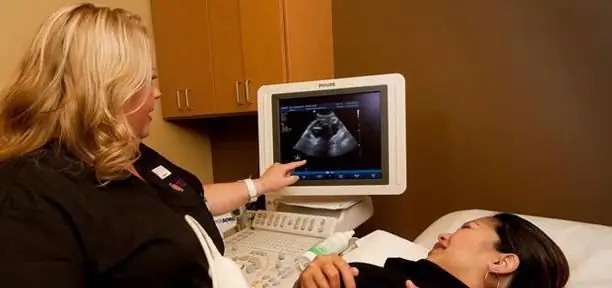
What to do if bleeding and pain accompany your pregnancy? On ultrasound in early pregnancy (the photo of the procedure is presented to your attention), everything will become clear. The specialist will determine the cause of your discomfort. Often, detachment of the fetal egg or insufficiency of the corpus luteum is diagnosed. With the timely detection of the pathology and its correction, the pregnancy continues to progress safely.
It is worth noting that the described symptoms may be a sign of an ectopic pregnancy, which was mentioned above. To clarify the diagnosis, it is necessary to do an ultrasound. In some cases, these symptoms still lead to termination of pregnancy. If in doubt, the doctor will prescribe you an additional study. The recommended interval between diagnostics should be at least two weeks.

In vitro fertilization
When in vitro fertilization, a woman must be assigned an ultrasound examination in the early stages. It is necessary to assess the condition of the genital organ. When replanting embryos, usually several embryos are selected. Diagnostics allowsdetermine how many embryos survived.
It is worth noting that you should not go for an ultrasound on your own in this situation. Contact your doctor. This specialist already knows all the features of your body and important nuances.
How is pregnancy diagnosed?
On an ultrasound scan in the early stages of pregnancy (the doctor will print the photo if you wish), the uterine cavity is examined. The specialist must determine the size of the reproductive organ. A comparison is made with the true terms (on a monthly basis). The condition of the ovaries is also assessed. One of them should contain a corpus luteum that supplies progesterone.
In the earliest stages, the doctor discovers the yolk sac near the embryo. This formation gradually decreases towards the end of the first trimester. Be sure to set the number of fruits and the place of their attachment. The duration of the diagnosis depends on the professionalism of the doctor and the operation of the equipment. On average, the diagnosis lasts 10-20 minutes. In the presence of pathologies, the specialist may need more time.

To sum up: the conclusion of the article
Is early ultrasound harmful? As you already understood, this question cannot be answered unambiguously. Diagnosis can bring both benefit and harm. It is necessary to consider each case individually, together with a specialist. Some women believe that ultrasonic waves can adversely affect the he alth of the fetus. However, this opinion is incorrect. If the doctor prescribes the described procedure for you, then it is necessary to carry it out. Be aware that ultrasound often reveals hidden problems.
In the absence of indications for diagnosis, you should not run for an ultrasound on your own. Excessive exposure to the uterus can lead to an increase in its tone. This consequence can be quite dangerous for the he alth and development of the embryo. If something is bothering you, be sure to consult a doctor. Do not prescribe a study yourself, especially since only a doctor can decipher the result.
Recommended:
How to distinguish pregnancy from ectopic pregnancy? Signs and symptoms of an ectopic pregnancy in the early stages

Pregnancy planning is a responsible business. And many women are thinking about how to understand that conception has occurred. Unfortunately, sometimes a pregnancy can be ectopic. This article will talk about how to recognize it in the early stages
Norm for screening ultrasound of the 1st trimester. Screening of the 1st trimester: terms, norms for ultrasound, ultrasound interpretation

Why is 1st trimester perinatal screening done? What indicators can be checked by ultrasound in the period of 10-14 weeks?
Ovarian pregnancy: causes of pathology, symptoms, diagnosis, ultrasound with photo, necessary treatment and possible consequences

Most modern women are familiar with the concept of "ectopic pregnancy", but not everyone knows where it can develop, what are its symptoms and possible consequences. What is ovarian pregnancy, its signs and methods of treatment
33 week of pregnancy: sensations, ultrasound, weight, height, development and photo of the fetus, examinations, recommendations

33-34 weeks of pregnancy - this is the period when a woman is overcome by excitement before the upcoming birth, and all sensations are noticeably aggravated. Almost all the thoughts of the future mother are occupied with the baby, worries about his he alth and a successful outcome of the pregnancy. All women are faced with the fact that by this time they think about the risks of preterm birth and begin to more carefully monitor their condition
What should be the discharge in early pregnancy?

Every woman has discharge in the early stages of pregnancy, which is a natural physiological process. In this regard, there should be no cause for concern. Normally, they should be white, but if a woman notices a different shade, then she should immediately visit a doctor for a consultation. It is better to play it safe once again than to worry about the consequences, among which there are very dangerous

