2026 Author: Priscilla Miln | [email protected]. Last modified: 2025-01-22 17:55:24
During pregnancy, the expectant mother should be regularly examined by a doctor. On average, a European woman visits a doctor 10-15 times during the entire prenatal period. Of course, the number of visits varies greatly depending on the individual course of pregnancy.
Most of the visits to the clinic are very conditional, but on them the couple can ask questions of interest, especially if the pregnancy is the first.

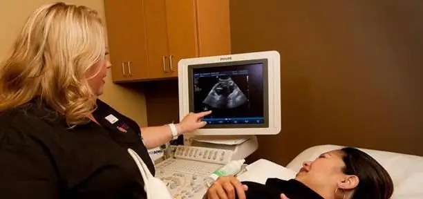
Among the routine procedures, there are some very exciting ones for those who are going to become parents in the near future. Of course, mom is really looking forward to meeting the baby when she can see him for the first time on the monitor during an ultrasound. As the embryo develops, the picture on the screen will become more and more informative.
Ultrasound at 12 weeks pregnant already shows a real tiny man. Further research will allow you to see the development of the crumbs and even draw a portrait of a new family member.
The safest method
It is believed that this method of diagnosis is the most harmless, and yet experts do not recommendabuse it on a whim, and go for examination only as directed by an obstetrician-gynecologist.
An extraordinary ultrasound is prescribed if the doctor is suspicious of the amount or structure of the amniotic fluid, the location or condition of the placenta, the length of the cervix, and the status of the myometrium uterus. All other examinations are carried out exclusively in accordance with the established calendar.
Ultrasound Calendar
The first ultrasound may be scheduled at 5-8 weeks, but more often this examination is skipped. A woman can not always immediately find out that she is expecting a child. If the study is done, then at this stage the very fact of the onset of conception, its estimated timing and the viability of the fetus are established. The photo shows only the presence of the embryo. In the period from 10 to 13 weeks, another ultrasound should be performed. The specialist uses a photo at week 12 to better examine the collar zone, as well as the attachment of the placenta and amniotic fluid parameters. At this stage, it is already possible to more accurately set the date of birth with an error of several days.

Between 20 and 24 weeks, another ultrasound examination is performed. Particular attention is paid to the correspondence of the activity and size of the baby, the state of the placenta and amniotic fluid. All measurements are compared with previous results obtained during ultrasound at 12 weeks of gestation.
The fourth study shows development over time compared to previous episodes. Immediately assess the fetoplacental and uteroplacental blood flow withusing the so-called Doppler method. The last sound examination is performed just before the appearance of an already formed baby. The doctor pays attention to the location and condition of the child, the position of the umbilical cord.
Photo to album
Many parents begin to form a photo album crumbs even before birth. Today, getting an ultrasound card is easy.

It is only worth notifying the doctor about this, and he will be happy to provide pictures. So you can collect a decent collection by the time of birth, with descriptions and annotations about the development, weight, condition of the child. The most interesting pictures are obtained at the last examination, when the baby's facial features are already formed. Then it will be interesting to compare ultrasound images and photos after birth.
Pregnancy monitoring
While external changes are not so noticeable, with the help of modern technology, the doctor can determine the size of the fetus, set the estimated time of delivery. All this is possible already with ultrasound at the 12th week of pregnancy.
The specialist analyzes the condition of the uterus, its tone, studies the location of the placenta, clearly establishes the physiological location of the fetus.
A pregnant woman is unlikely to be able to independently understand the image on the screen, so you should ask for an explanation from the operator. At this stage, the doctor is guided by tabular indicators and draws conclusions about the course of gestation, comparing them with the results of ultrasound at week 12. Pregnancy rates are determined based on observations of the general course of mostpregnancies.
Who will the stork bring
There are a huge number of ways to find out what gender the unborn baby will have. On ultrasound at 12 weeks, you can already find out.

Most of the parents after this procedure get the answer to the long-awaited question. However, sometimes it is not possible to establish the sex until the child's birthday, although the reproductive system has already been formed. People say that the baby will be shy if he constantly hides the bottom of the tummy from the apparatus. How to be in this case?
There is a study that determines who will be born based on the rhythm of the heart. It is believed that in girls it is more than 140 beats per minute, in boys it is less. This can be found out from the ultrasound operator at the 12th week of pregnancy. However, this is more likely a speculative observation than a fact confirmed by medicine. The probability of a match is 50%, the same as the probability of just guessing. Another popular way to guess the sex of a child is to look at the shape of the abdomen. The higher position of the fetus in the womb is believed to mean a girl, and the "lower" belly means a boy. However, gynecologists say that another factor affects the position of the fetus and, as a result, the shape of the abdomen. We are talking about the width of the hips, the narrower the hips - the sharper the shape of the abdomen will be.
The eating habits of the expectant mother also indicate the sex of the child. It is believed that a preference for sweets indicates a developing girl, and meat - a boy.
If a pregnant woman seems to fade while carrying a child, then a daughter will be born. They say that the daughter eats her mother'sbeauty. At the same time, the boy in the womb reveals the attractiveness and brightness of the woman's appearance.
Inside work
At 12 weeks, at the end of the first trimester, the baby continues to grow rapidly.

The legs and arms grow, the abdominal cavity is formed, in which the intestines are distributed. The rudiments of downy hairs are born: future eyebrows, eyelashes, hairs on the cheeks and above the upper lip are laid. Fingerprints emerge.
At this point, the immune system is actively differentiating. The glands begin to produce their own hormones.
The baby actively somersaults, wrinkles his lips, swallows liquid, blinks his eyelids, turns his head and sucks his finger. At this time, you can hear the baby's heartbeat using a Doppler device.
In the position of a future mother
The expectant mother's uterus is growing. During the moment of pregnancy, the uterus can stretch from 10 ml to 10 liters, from 70 g to 1100 g. The breast becomes more sensitive, its size increases. Weight gain by this period is 2 - 3.5 kg. A woman is recommended to have an ultrasound at the 12th week of pregnancy of the internal organs.

Red pigment spots, itching may appear on the skin, indicating overstretching of the skin. It is at this stage that it is important to begin to properly moisturize the epidermis with the help of special creams. Do not be afraid of the appearance of a dark strip near the navel, all this will pass after childbirth. The heart of the expectant mother begins to beat faster in order to pump moreamount of blood. But urination becomes less frequent compared to the first three months.
A woman is already fully accustomed to her new role and is easier to endure the chores that the intestines can deliver. Flatulence and constipation in most cases are easily regulated by nutrition. In the case of individual characteristics or pregnancy with twins, toxicosis may persist. There are other features of multiple pregnancy.
Carrying twins
Just a few generations ago, the arrival of twins was often a big surprise. However, thanks to the use of ultrasound in medicine, multiple pregnancy can be diagnosed as early as 5 weeks.

More often the fact is established when the first ultrasound is done at 12 weeks. The realization that two children will be born at once is important for the psychological preparation of parents, as well as for the necessary preparations, such as buying cradles and special strollers designs.
In addition, the examination will help to do everything possible to prevent complications that occur more often in multiple pregnancies.
These include preterm birth and gestational diabetes. It is also important that midwives and birth attendants are aware of this. Carrying twins is more difficult, so you need to visit the doctor and do control tests more often. Thus, the first ultrasound of the baby at 12 weeks can give parents very important knowledge and prepare them for the upcoming tasks.
More accurate than ultrasound
For more information aboutduring pregnancy, it is worth turning to a deeper study than ultrasound. Screening at week 12 uses data obtained from blood biochemistry. Two markers are analyzed: free b-hCG and PAPP-A. That is why the procedure is also called a double test.
Doctors recommend conducting an examination three times during the entire pregnancy.
Ultrasound during screening is carried out in more detail and makes it possible to control the "collar zone" in the fetus, which, in turn, turn, eliminates gross defects or anomalies. The zone is located between the soft tissues and the skin, where fluid accumulates. In the course of pregnancy, the space is constantly changing, and in order for these temporal markers to provide the necessary information, screening must be done clearly at the time prescribed by the doctor. Such a study can only be done by a highly qualified operator, since the subjectivity of the data is quite high.

Blood test results are also designed to rule out abnormalities in the fetus. An increase in b-hCG by half raises the suspicion that the fetus has trisomy 21 (Down's syndrome), and a decrease - Edward's syndrome. But all this is just an assumption, which, of course, devalues the results.
Pros and cons
Ultrasound at 12 weeks or a little earlier - usually 8-12 weeks - this is additional information for the therapist about the exact date of conception and the activity of the embryo. The expectant mother does all the tests of her own free will, guided by the interests of the baby's he alth.
Recommended:
Fetal size at 8 weeks of gestation: stages of development, sensations, photos from ultrasound

Having learned about her new status, a woman tries to listen to the slightest changes in her state of he alth. Since her feelings change every week, she needs to understand which symptoms are normal and for how long, and which ones are a signal to see a doctor
HCG at 5 weeks of gestation: decoding analysis, norms, pathology and advice from gynecologists

For any woman, a long-awaited pregnancy will be a great joy in her life, and being pregnant, she takes care of the he alth of the unborn baby in the womb. Throughout the trimesters of gestation, all women are assigned a large number of various studies to make sure that everything is in order with the fetus inside. In this article, we will take a closer look at what hCG should be at the 5th week of pregnancy, what this analysis is
Norm for screening ultrasound of the 1st trimester. Screening of the 1st trimester: terms, norms for ultrasound, ultrasound interpretation

Why is 1st trimester perinatal screening done? What indicators can be checked by ultrasound in the period of 10-14 weeks?
Delivery at 37 weeks of gestation: the opinion of doctors. How to induce labor at 37 weeks?

Pregnancy is a very responsible period for every woman. At this time, the body of your crumbs is formed and develops. In many ways, the he alth of the future depends on the course of pregnancy
Fetal CTG is the norm. Fetal CTG is normal at 36 weeks. How to decipher fetal CTG

Every expectant mother dreams of having a he althy baby, so during pregnancy she worries about how her child develops, is everything okay with him. Today, there are methods that allow a fairly reliable assessment of the condition of the fetus. One of them, namely cardiotocography (CTG), will be discussed in this article

