2026 Author: Priscilla Miln | miln@babymagazinclub.com. Last modified: 2025-01-22 17:55:27
Every woman expecting offspring worries about whether her pregnancy is developing correctly. To ensure full control, it is recommended to visit a gynecologist in a timely manner. He will be able to correctly determine the size of the fetus by weeks of pregnancy, as well as identify certain deviations. For accurate diagnosis, modern ultrasound methods are used, which allow you to compare the data obtained with the norms of fetal development.
Why you need to know the sizes

For every expectant mother, doctors determine the size of the fetus by weeks of pregnancy, as well as its weight. What is it for? The doctor can notice deviations in time and take action, and the date of birth will be set more accurately. If these indicators are monitored regularly, it is easy to recognize a missed pregnancy, which can threaten a woman's he alth.
Baby weight at different stages of developmentalso indicates how the birth will take place. If the fetus is too large, then a caesarean section may be required. If the baby is too small, it may happen that he needs first aid immediately after birth.
Fetal size by week of pregnancy by ultrasound

Measuring the size of the fetus using ultrasound is called fetometry. It is carried out in two ways:
- A special small probe is inserted into the inside of the vagina (vaginal method).
- The sensor is driven along the surface of the abdomen (abdominal method).
In the first trimester of pregnancy, the following are considered the main indicators of size:
- Fertilized egg. The size of the cavity in which the embryo develops is measured.
- Biparietal distance. The gap between the right and left temporal bone.
- Coccyx-parietal size. This is the distance from the tailbone to the top of the head.
In the 2nd-3rd trimester there are more indicators, these are:
- Fetal growth.
- Thigh bone length.
- Biparietal head size.
- Chest (diameter).
- Circumference and girth of the abdomen.
- Length of the humerus.
- The distance between the forehead and the back of the head.
Deviations from the norm
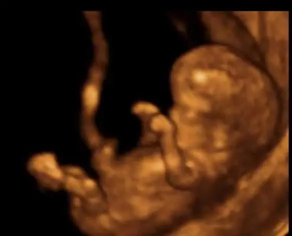
All babies can develop differently, with jumps in periods. Doctors are guided by average indicators. So, the size of the fetus at 6 weeks of pregnancy (photo is presented) for one mother may be slightly larger, for the other - slightly smaller. itIt also depends on the genetics of the parents. For the entire period of development, measurement of dimensions is carried out several times. Pathology is the case if several indicators at once are much different from the average norm.
Weight gain or loss
What is the size of the fetus by week of pregnancy? This is clearly visible on the ultrasound photo. If the fetus is too small, you should pay attention to the parents themselves, perhaps they both have a small complexion. Another reason may indicate bad habits of the mother (smoking, drinking alcohol); for the use of antibiotics. The fetus may grow slowly due to poor oxygen supply. In this case, the mother is advised to immediately give up alcohol and smoking, start eating well and stop taking antibiotics. Too rapid weight gain in the fetus may indicate that a woman is abusing fatty foods. Sometimes the cause of a lot of weight is diabetes, which mommy suffers from.
Decrease or increase in CTE (indicates the coccygeal-parietal size)
When measuring the size of the fetus by week of pregnancy, the KTP indicator is used. It is important up to 13 weeks. The rapidly growing KTR numbers indicate the factor that the fetus will grow very large in the future (4 or more kg). The doctor in this case does not recommend taking multivitamin complexes that speed up the metabolism. With a low CTE, there are suspicions of the following deviations:
- Hormonal deficiency (appointment of "Dufaston" and "Utrozhestan").
- Suspicion of infection (an additionalresearch and then treatment).
- Genetic disorders in the development of the fetus (for example, Down syndrome, etc.).
- Diseases of internal organs in a woman (examination is scheduled).
- Death of the fetus (emergency surgery to remove the embryo).
Decrease or increase in BDP (indicates biparietal head size)

If BDP is too low, this may indicate that there is a developmental delay. There is a chance that after birth, the baby will be identified with various congenital malformations. Elevated BDP indicates the possibility of dropsy or hydrocephalus. The fetus in this case may die if fluid accumulates in the brain cavity.
Size by week of pregnancy (weeks 1-10)
1 week. This reference point has several concepts. If we talk about the obstetric week, then the first day of menstruation, which was recorded for the last time, is taken into account, and after that the woman had unprotected sex. If you calculate the moment of conception by day, you get the third obstetric week. When taking into account the date of the delay of menstruation, you can get the fifth week. In gynecology, following the development of pregnancy, they rely on obstetric terms. The first week is not characterized by any special signs. This time is considered the beginning of the menstrual cycle. At the same time, the level of hCG at this moment is normal.
2 week. The obstetric week is characterized by the maturation of the zygote, with favorable contact, it will develop into pregnancy. The fertilized egg will attach itself to the wall of the uterus. Evidence of this may be a discharge similar to egg white. Small blood impurities may appear due to the attachment of the egg. This is the norm.
3 week. At this time, we can say for sure that the conception has happened. The size of the fetus is too small: 0.15-0.20 mm long and weighs 2-3 μg. In case of unsuccessful fertilization, if the egg does not attach, the onset of menstruation is possible a little earlier than the calendar.

4 week. The development of the embryo is too active. A woman feels the first signs, changes in the body. The mammary glands begin to swell, the nipples are too sensitive. Delayed menses. There may be scanty bleeding. During this period, the risks of anomalies in the development of the fetus are high, if a woman has increased physical activity, has infections, fever, she abuses alcohol. The level of hCG in the blood rises. On ultrasound, the corpus luteum is determined, which nourishes the embryo and is involved in the active production of progesterone (pregnancy hormone). The fruit is already 5 mm long.
5 week. The length of the fetus is now already 4-7 mm, and its weight is 3.5 g. The formation of the rudiments of the limbs, auricles, eyes, slits of the mouth and nose, and some glands begins. The size of the uterus changes. At this time, an ultrasound can already be seen - a singleton or multiple pregnancy is developing. Establish KTP, the size of the fetal bladder, the growth of the fetus.
6 weeks pregnant. The size of the fetus becomes larger, its length is 4-9 mm, while the weight is approximately 4.5 g. Motherfeel changes in the body. The uterus increases to the size of a plum. At the 6th week of pregnancy, the size of the fetus, as well as the number of fetal sacs, is clearly visible on ultrasound. Small tubercles are noticeable, limbs will form in these places. On special devices it is already possible to listen to the heartbeat.

7 weeks pregnant. The size of the fetus already has a length of 13 mm. The heart is divided into four chambers and blood vessels are formed. Internal systems and organs develop. The embryo begins to straighten up a little. The brain is actively developing. The umbilical cord is already fully formed.
8 week. The length of the fetus is already 14-22 mm. Slowly, he starts to move. The face begins to take on a human shape. The laying of systems and organs is being completed, many are already starting to function. Genital organs and optic nerves are born.

9 week. Growth is 22-30 mm, with a fetal weight of 2 g. The cerebellum, the middle layer of the adrenal glands, the pituitary gland, the genital organs, and the lymph nodes are formed. Limbs begin to move, muscles form. Ability to urinate.
10 week. The first critical stage of development is coming to an end. The weight of the baby is 5 g with growth reaches 40 mm. Heartbeat 150 beats / min. You can see the fingers and joints of the limbs. The organs of the gastrointestinal tract are completing their formation. The foundations of the teeth are being laid. At this time, calcium intake is especially important for mothers.
11-20 development weeks
11 weeks pregnant. The size of the fetus in length is already 5 cm, in weight - 8 g. The embryo at this time can already be called a fetus. The blood vessels are formed, and the heart works fully. In the intestines, the first movements are observed, which are similar to peristalsis. The reproductive organs of the fetus continue to develop. There is a sense of smell, eye color. Fingers and palms acquire the ability to feel.
12 weeks pregnant. The size of the fetus is already within 6-8 cm. Nails form on the fingers. The gastrointestinal tract is completing its formation. The immune system develops. At the 12th week of pregnancy, the size of the fetus becomes larger, the uterus increases. Mommy feels her tummy start to grow.
13 week. The beginning of the second trimester of pregnancy. Fetal growth is 8 cm with a weight of 15-25 g. Milk teeth are laid, the formation of muscle and bone tissue, the digestive system, and genital organs continues.
14 weeks pregnant. The size of the fetus already reaches 10 cm in some cases, while its weight is 40 g. A skeleton and ribs are formed. The main organs and systems are already fully formed. The baby already has a blood type and his own Rh factor.
15 weeks pregnant. The size of the fetus reaches 10 cm with a weight of 70 g. The cerebral cortex is formed. The work of the endocrine system, sweat and sebaceous glands is activated. Formed taste receptors, respiratory movements. In the uterine cavity, the baby moves freely.
16 week. During this period, the growth of the fetus is 11 cm, while the weight is 120 g. The head rotates freely. Ears and eyes rise up. The liver begins to work. Formed the composition of the blood.
17a week. The immune system begins to work. The baby can already to some extent protect itself from infections that threaten the mother's body. A fatty layer is formed. Height is 13 cm with a weight of 140 g. The child begins to feel emotions, hear sounds from the outside. During this period, it is extremely important to establish contact with the baby.
18 week. Middle of the second trimester. The limbs are fully formed, there are already prints on the fingers. The rudiments of molars appear. There is a further development of the immune system, brain and adipose tissue. Hearing intensifies, there is already a reaction to light. Fruit weight 200 g, height 14 cm.
19 week. A strong leap in development. The movements are more perfect. The baby is already freely rotated or held in any position. Lubrication appears. Height - 15 cm, weight 250 g.
20 weeks pregnant. The size of the fetus at this time is 25 cm, and the weight is 340 g. The baby is fully formed. The heartbeat can already be heard with a stethoscope. Mom begins to acutely feel the movements of the fetus.
21-30 Development Weeks
21 weeks pregnant. The size of the fetus with a height of 27 cm already weighs 360 g. There is still enough space for active movement in the uterus. The fetus begins to swallow amniotic fluid. Muscle and bone tissue is already strengthened. The spleen begins to work.
22 week. There is a significant weight gain - up to 500 g. Growth also reaches 28 cm. Even if born, the fetus may already be viable. The spine and brain are already fully formed. The heart is growing. Reflexes are improving.
23a week. The fruit is formed. The digestive system is functioning. Adipose tissue grows. The genital organs are clearly differentiated. Baby weighs 500g and is 29cm tall.
24 week. Weight 600 g, and height - 30 cm. There is more and more adipose tissue. The production of growth hormone begins. Sense organs and reflexes are improved. There is already a pattern of sleep and wakefulness. The baby reacts sharply to the emotions of the mother.
25 week. The baby has now grown to 34.5 cm, weight is 700 g. He looks more and more like a newborn. Strongly developed sense of smell, emotions. The lungs prepare for independent breathing. The vagina and testicles appear.

26 week. Individuality emerges. Eyes open. The baby recognizes familiar voices. Bone tissue is strengthened. The lungs are formed. Various hormones are produced. The weight of the baby is 750 g, its length is 36.5 cm.
27 week. By this time, the growth of the fetus is more active. Its weight already reaches 900 g. Metabolic processes in the body are developing. The endocrine system and the brain are actively working. Subcutaneous fat becomes more and more. Mommy increasingly feels the movements of the baby.
28 week. At this period, the baby is already gaining his first 1 kg. Height is 38.5 cm. The space inside the uterus becomes much smaller, but this does not affect development in any way.
29 week. The baby's body begins to prepare for the birth of the world. The work of the immune system, thermoregulation is debugged. The composition of the blood is stabilized. The digestive system is ready. The skin becomes wrinkle-free, lighter. Look alreadyfocused. Strengthens muscle tissue.
30 week. The kid gained 1.5 kg. The nervous system is included in the work. Iron accumulates in the liver. The eyes are often open. The fetus, as a rule, is already in the position that should be at birth.
31-40 weeks pregnant
31 week. The baby already weighs more than 1.5 kg. The production of surfactant continues. The liver cleanses the blood. Communication between the brain and peripheral nerve cells has already been established. If the baby touches the cornea, then the eyes close. The development calendar is coming to an end.
32 week. Active development continues. Systems and organs are fully operational. Appearance takes on the appearance of a baby. Fluff disappears. The skull remains soft, the baby is in the prenatal position.
33 week. Weight normally reaches 2 kg. Build muscle and subcutaneous fat. The baby can express emotions. The kidneys are getting ready for filtering work.
34 week. Now the development of the fetus is almost complete. There are training of the digestive tract. Individual facial features are becoming more distinct.
35 week. In this period, the organs have already developed. Basically, a set of muscle and fat layers is being carried out. Weekly gain up to 220g
36 week. The body is improving. The work of vital systems is being debugged. Iron continues to accumulate in the liver. The baby actively sucks his finger, so the preparation for sucking the breast begins. Normal - cephalic presentation of the fetus.
37 week. The fruit is finally formed. Intestinal peristalsis is activated. Heat exchange processes are established. Lungsripe. Weekly gains in height and weight.

38 week. The baby is now ready to be born. The skin becomes pink. In a male child, the testicles descend into the scrotum.
39 week. The organs and systems of the baby are ready for independent work. Reaction to light and sounds is well developed. The original lubrication is no longer on the skin.
40 week. The approximate height of a newborn is 54 cm, while the weight is from 3 to 3.5 kg. The baby will be born soon, the formation of the fetus is fully completed.
It is important for every mother to know how a child develops inside her. This will allow you to correctly respond to changes in your own body and, as soon as necessary, turn to a gynecologist in time. Observations are important throughout the pregnancy.
Recommended:
Can an ultrasound not show pregnancy? Fetal size by week of pregnancy

There are times when women find out they're pregnant when they're well into their term. There are quite a few ways to confirm a special situation using the analysis of hCG, various tests. But sometimes, the listed methods do not always carry reliable information. Can an ultrasound not show pregnancy? We'll talk about it in this article
Light fetus - a pathology or a feature of the constitution? The norm of fetal weight by week

A child is real happiness and real work, from which you can’t run away. Many expectant mothers are afraid of complications, because everyone wants to give birth to a he althy baby. But a small fetus is not a sentence, children are born he althy
Size of the abdomen during pregnancy: abdominal circumference by week, fetal development, photo

The size of the abdomen during pregnancy is measured in order to monitor the course of bearing the baby. It helps to determine how the pregnancy is proceeding, whether there are any pathologies. But do not forget about the individual characteristics of the body
What happens in the 12th week of pregnancy. 12 weeks pregnant: fetal size, baby gender, ultrasound picture

12 weeks pregnant is the final stage of the first trimester. During this time, a little man has already developed from a cell that is visible under a microscope, capable of making some movements
Should I do an ultrasound in early pregnancy? Pregnancy on ultrasound in early pregnancy (photo)

Ultrasound came into medicine about 50 years ago. Then this method was used only in exceptional cases. Now, ultrasound machines are in every medical institution. They are used to diagnose the patient's condition, to exclude incorrect diagnoses. Gynecologists also send the patient for ultrasound in early pregnancy

