2026 Author: Priscilla Miln | miln@babymagazinclub.com. Last modified: 2025-01-22 17:55:13
Ultrasound diagnosis is included in the list of procedures provided by the obstetric pregnancy support program. This is the most informative and safe method, which currently has no alternative. By learning about the condition of the developing fetus, parents can satisfy their curiosity and ensure that the baby is born he althy.
Characteristic features of 3D ultrasound

Like standard diagnostics, 3D examination is based on the penetrating power of ultrasonic waves. A two-dimensional image allows the doctor to obtain valuable information and make a medical conclusion, but for future parents it is of little interest.
The term "3D" indicates that the image conveys not only the width and height, but also the depth of the object being studied. Thus, a color image of the embryo is displayed on the screen, which can be written to a disk and left as a memory of the first acquaintance with the child. It is important to understand at what time it is better to do 3D ultrasound andPlease note that this method is not a replacement for standard diagnostics. Volumetric scanning allows you to study in detail the external structure of the fetus, and conventional ultrasound reveals pathologies in the development of internal organs.
Advantages and disadvantages of the method
3D ultrasound is becoming more and more popular. This is due to its undeniable advantages:
- large, compared to conventional scanning, information content;
- accurate definition of external developmental pathologies such as congenital cleft lip and palate, better known as cleft lip and palate;
- obtaining a three-dimensional image of the internal organs, allowing you to track possible deviations at an early stage;
- opportunity to assess the functioning of the cardiovascular system of the embryo;
- assessment of the emotional comfort of the child by his facial expressions: a smile indicates good he alth, and a grimace of pain or lethargy indicates the presence of certain disorders;
- accurate sex determination in any position of the fetus;
- in multiple pregnancies, only 3D ultrasound can provide reliable information about the development of children;
- photo allows parents to take a good look at the baby, see his smile.
The procedure also has some drawbacks:
- comparatively high cost;
- increased research time;
- if a woman gains excess weight during pregnancy, image quality will be reduced;
- if the baby turns his back, you won't be able to see his face.
Does the procedure harm the baby

Ultrasonic radiation can penetrate the human body. For this reason, many expectant mothers are worried that the baby will be harmed. It should be noted that data on the destructive effect of ultrasound were obtained as a result of the use of amplified sound waves. This power is not used during the study.
Specialists say that an ultrasound scan, carried out in accordance with all the rules, does not harm either the mother or the embryo. According to WHO, it is considered safe to undergo four ultrasound examinations, while for a period of less than 10 weeks, the procedure should not be performed unless absolutely necessary. In many modern clinics, 3D ultrasound is not done without an appropriate referral. Experts refer to the conclusion provided by the American agency FDA, which does not allow for the possibility of additional ultrasound sessions solely for the peace of mind of parents. In this case, you need to contact the doctor in charge of the pregnancy, who will be able to recommend at what time it is better to do 3D ultrasound, and write out a referral.
Destination of scanning depending on the term

In the prenatal period, ultrasound monitoring is carried out as planned. The terms are determined by the obstetrician-gynecologist observing the woman, guided by medical standards:
- From 5 to 8 weeks, you can establish the presence of pregnancy, make sure that the fetal eggfirmly attached to the wall of the uterus, and the embryo is viable.
- From 10 to 14 weeks, ultrasound allows you to determine the exact date of conception, designate the expected date of birth and obtain data on the compliance of the child's development with the norms.
- From the 16th to the 23rd week, a specialist can exclude the presence of malformations in the embryo, monitor the formation of vital organs, check whether the size of the baby corresponds to the established deadlines.
- From 30 to 32 weeks, the procedure is carried out in combination with Doppler ultrasound. This allows you to make a conclusion about the overall development of the fetus and its motor activity.
- Monitoring before childbirth is necessary to detect presentation, control the he alth of the embryo and its body weight.
When is it better to do 3D ultrasound during pregnancy

This question worries young parents, especially those who are expecting their first baby. As for how long it is better to do a 3D ultrasound with a photo during pregnancy, experts are of the same opinion. It is best to study the three-dimensional image of the child from 24 to 28 weeks of the prenatal period. This period is considered the most appropriate for the following reasons:
- fetal organs are already formed and moderate radiation will not adversely affect their development;
- it is possible to examine the baby's facial features, determine his gender, find out if the limbs are correctly formed;
- due to its small size, the embryo can still move freely in the uterus, so observehis activities are more interesting;
- baby rolls over easily, even if the child was initially positioned with his back to the sensor, there is a chance that he will take a better position during the examination;
- a woman already feels the fetus moving, and can choose the time of day when the baby is most active.
Medical indications
Patients often ask a pregnancy specialist when it is better to do a 3D ultrasound of the fetus. The optimal time for the procedure is selected taking into account medical indications:
- risk of developmental pathologies in the embryo;
- genetic predisposition to dangerous diseases;
- detection of fetal defects;
- surrogate pregnancy;
- passing the IVF procedure;
- multiple pregnancy;
- determining the sex of the child.

Many institutions allow 3D diagnostics without a medical referral so that parents can see their baby for the first time. It is believed that this allows future fathers to better get closer to the baby. It is important to independently monitor that the frequency of studies does not exceed the norm.
Necessary preparations
Before undergoing the procedure, you need to consult with a specialist to find out at what time it is better to do 3D ultrasound during pregnancy with recording to a disk. This will make the diagnosis as informative as possible. No special preparation is required for the study. Since after 20 weeks of gestationThe baby's uterus occupies a significant part of the abdominal cavity, the fullness of the bladder will not interfere with visualization.
To facilitate the work of the doctor, it is advisable to avoid eating foods that can cause gas accumulation 1-2 days before the procedure:
- legumes;
- black bread;
- fresh fruit;
- cabbage;
- carbonated drinks.
Ideally, before an ultrasound, you should empty your bowels naturally. With enemas, you need to be careful, squeezing the uterus can cause premature birth. Severe bloating may be a reason to postpone the procedure and consult a doctor who will tell you how to improve digestion and how long you can do 3D ultrasound.
Diagnostics

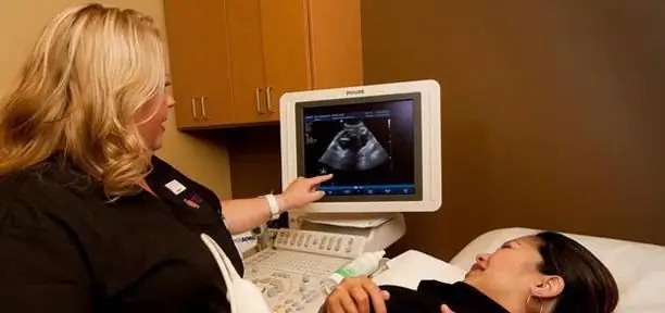
A patient who has decided on the date for 3D ultrasound during pregnancy must sign up for a diagnosis. The procedure is carried out in the same way as a two-dimensional scan - using a standard probe. To facilitate the sliding of the device, the woman's stomach is lubricated with a special gel. Thanks to the special principle of moving the device, a computer can build a three-dimensional image based on the received data. Scanning takes 5-10 minutes. It takes another 30-40 minutes to process the result.
The study will be informative only if the woman managed to find out at what time it is better to do 3D ultrasound. You should also understand that the baby can turn his head to the sensor.
Where can I get a 3D ultrasound
The necessary equipment can be found in gynecology and obstetrics centers, as well as in private clinics. Patients of the antenatal clinic are offered only two-dimensional diagnostics. When choosing an organization, many expectant mothers go to the forums and read reviews on 3D ultrasound during pregnancy. At what time is it better to do the procedure, the fair sex asks the doctor observing them.
The procedure may be included in the pregnancy management program offered by specialized clinics. In addition, the service can be paid separately. In this case, its cost will be from 1.5 to 4 thousand rubles. The price includes recording data on digital media.
Patient testimonials

The 3D scanning procedure is considered quite popular and more and more women are interested in how long it is better to do 3D ultrasound during pregnancy. Reviews of pregnant women indicate that the procedure guarantees future parents a lot of positive emotions. Especially if the study is carried out for a period of 24 weeks. At this time, the baby is already quite developed and you can examine in detail the features of his face, fingers and toes. Expectant mothers were especially impressed by the serene smile of the crumbs.
For women who care about the he alth of the child, it is important not only to get a photo as a keepsake, but also to make sure that the baby has no pathologies. Experienced specialists, having studied the data obtained, immediately call the approximate date of birth, explain how the fetus develops, whether there is a growth lag. Patients note that if ultrasoundis carried out in a free direction, the emphasis is on the features of the formation of organs and systems. Women who underwent the procedure in a private clinic were able not only to obtain information about the he alth of the crumbs, but also to observe him. And photos recorded on digital media can be shown to all relatives and friends.
Three-dimensional ultrasound is not dangerous to he alth. But you should not exceed the number of procedures recommended by your doctor.
Recommended:
4D ultrasound during pregnancy: results, photos, reviews

Today, many clinics offer a medical service such as "4D Ultrasound for Pregnant Women". What is such a diagnostic procedure, why is it carried out and how safe is it, we will tell in our material. We will also share feedback from doctors and patients about this study
Norm for screening ultrasound of the 1st trimester. Screening of the 1st trimester: terms, norms for ultrasound, ultrasound interpretation

Why is 1st trimester perinatal screening done? What indicators can be checked by ultrasound in the period of 10-14 weeks?
Who is better a cat or a dog? Who is better to start: pros and cons

The article discusses the issue of choosing an animal, talks about both the problems that owners may face and the joys of living together
JBL headphones sound better and better than expected

JBL headphones are a product that is known and in demand in all corners of the planet. Many still only dream of such headphones, but many have already appreciated their advantages, and not a single buyer will regret such an investment, because such “ears” are really worth the money
Jelqing: reviews and results. The benefits and harms of jelqing

Surprisingly, not only women are unhappy with their bodies. As it turned out, a large number of men suffer because of the modest size of their main advantage. And although modern cosmetology and medicine know how to help them, the solution to this problem is very non-standard - jelqing. Reviews and results of using this technique, we will consider in more detail

