2026 Author: Priscilla Miln | [email protected]. Last modified: 2025-01-22 17:55:21
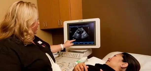
Ultrasound examination (ultrasound) is one of the main procedures performed during pregnancy. This is not only a necessary medical event that allows you to identify possible pathologies in the development of the fetus in the early stages, but also an exciting event for expectant mothers and fathers. This is a kind of acquaintance with your future baby. Despite the fact that ultrasound has been used in obstetrics not so long ago - since the 60s of the last century, doctors have accumulated vast experience in using this research method over this period of time. Ultrasound scanners are constantly being improved, and today it is possible to do 3D ultrasound during pregnancy. It is this type of research that will be discussed in this article.

What is the difference between conventional ultrasound and 3D ultrasound?
Two-dimensional ultrasound shows the image of tissue sections of the area affected by ultrasound. With 3D ultrasound, the image on the monitor screen looks three-dimensional and color. In addition, such a picture makes it possible to examine in detail the appearance of the baby and even see who he looks more like. With the help of such a study, you can determine:
- facial anomalies (cleft lip, cleft palate);
- developmental pathologies of the nervous system;
- nasal bone development and neck crease thickness;
- congenital heart defects.
How 3D ultrasound works
The method of such a study is not fundamentally different from conventional ultrasound, which uses the penetrating property of ultrasound and its ability to spread differently, depending on the composition and density of the medium, in the tissues of the body. However, the image obtained with a classic ultrasound is completely incomprehensible to non-professionals, and future parents can only detect large bones and a child's spine with the help of a doctor. With 3D ultrasound, the picture resembles an ordinary photograph, and happy mothers and fathers can see the baby’s face and even count fingers.

Benefits of 3D Ultrasound
Three-dimensional ultrasound examination, in addition to the great joy experienced by future parents, makes it possible to obtain more accurate and complete information about the course of pregnancy and the condition of the fetus. This procedure is especially indicated if there are any suspicions of developmental pathologies, as it makes it possible to identify deviations from the normal values of certain indicators at an earlier date.
This procedure allows you to examine various organs. With the help of a three-dimensional image, you can get an accurate picture of the state of the external and internal organs of the baby. The 3D study also allowslook at the facial expressions of the crumbs. This allows you to see what emotions he is experiencing: upset, smiling, apathetic. It has long been known that positive emotions allow the fetus to develop properly. But bad ones can indicate serious problems. For example, with asphyxia (insufficient oxygen supply), the baby has an apathetic, depressed state. If the child's face is distorted with pain, then this may indicate an abnormal development of the internal organs, which causes pain.
It is also very important that the future dad be present at the 3D ultrasound procedure. This will help him quickly adapt to the role of a father. Photo 3D ultrasound, if desired, can be the first picture in the album of the future baby.

At what stage of pregnancy should I do a 3D ultrasound?
Most parents want to get the very first touching photo of their baby as soon as possible. However, this should not be done before 18-20 weeks. At an early date, it will still be impossible to see something. It is possible to more accurately determine both the sex of the child and possible developmental deviations if the pregnancy is 20 weeks. 3D ultrasound in this case will be more informative. 3D examination allows you to view facial structures, head, back.
You should also know that every day the baby is getting closer to being in mom's tummy, and in the later weeks it is also not recommended to do 3D ultrasound. 32 weeks is the deadline for this procedure, since in the last weeks of pregnancy, as in the case of a two-dimensional ultrasound, it is more difficult for a doctor to getreliable information and high-quality image.

What should future parents know?
- Almost twice as long as a 2D study takes a 3D study. Therefore, you should be prepared for the fact that the 3D ultrasound procedure will last at least 30-40 minutes.
- Reviews of women who used this research method indicate that quite often the baby does not want to be "photographed" and simply turns his back. In this case, the procedure, of course, will not be as informative as we would like.
- You should also be aware that the procedure for three-dimensional examination of the fetus is not cheap. Its cost ranges from 1500-2500 rubles.
Is 3D ultrasound dangerous?
Whether or not ultrasound during pregnancy is safe is not completely known. But 3D ultrasound in terms of radiation strength is no different from a two-dimensional study. Therefore, the question is not about the method of obtaining a stereo image, but about ultrasound as such. To date, it has already been established that ultrasound is in no way associated with an increase in the frequency of congenital pathologies and pregnancy anomalies. That is why ultrasound is one of the mandatory procedures that pregnant women undergo.
However, there is such a medical term as long-term effects. This concept refers to complications that may occur only after a few years or even decades. It is these long-term effectsthe use of ultrasound during pregnancy has not been determined. No one can say for sure what will happen to these children in 10, 30, 50 years.

Do or not 3D ultrasound?
As mentioned above, the disadvantages of a three-dimensional study are its high cost and long procedure time. Trying to get the best shot, doctors often increase the power of the device during the study, and this can adversely affect the child. Because the possible effects of 3D testing have not been fully determined, many practitioners strongly recommend limiting its use to pregnant women.
In addition, the image may not be clear and of good quality when:
- proximity of the fetal head to the placenta;
- low amniotic fluid;
-
overweight pregnant.

3d ultrasound 32 weeks
What is 4D (four-dimensional) ultrasound?
This is the same as the 3D study, but it differs in that the length, height and depth of the picture are complemented by time. A 4D image, unlike a static 3D image, allows you to see the movement of an object in real time. This makes it possible for future parents to record what is happening on the monitor screen on various media.
Of course, the temptation to see your baby before birth is great enough. But future mothers should know that you should not abuse ultrasound. ultrasoundshould be an exclusively planned procedure, and not be conditioned by the desire of a woman to take another look at her baby. Well, whether to do a three-dimensional study or stop at the usual two-dimensional one, only future parents should decide.
Recommended:
"Cycloferon" during pregnancy - is it possible or not? Instructions for use of the drug during pregnancy

The use of "Cycloferon" during pregnancy in the early stages helps to get rid of the symptoms of viral and infectious disorders. Human immunity is activated, a stable antimicrobial effect occurs. Tumor formation in the body slows down, autoimmune reactions are restrained, pain symptoms go away
"Sinupret" during pregnancy in the 3rd trimester. Instructions for use of the drug during pregnancy

Infection and inflammation are more pronounced when the body is weakened, so experts choose safe medicines. Used "Sinupret" during pregnancy. The 3rd trimester passes without serious complications if the infection can be overcome on time with the help of this medicine
Cutting pain in the lower abdomen during pregnancy: causes. Drawing pain during pregnancy

During the period of bearing a child, a woman becomes more sensitive and attentive to her he alth and well-being. However, this does not save many expectant mothers from pain
Norm for screening ultrasound of the 1st trimester. Screening of the 1st trimester: terms, norms for ultrasound, ultrasound interpretation

Why is 1st trimester perinatal screening done? What indicators can be checked by ultrasound in the period of 10-14 weeks?
Should I do an ultrasound in early pregnancy? Pregnancy on ultrasound in early pregnancy (photo)

Ultrasound came into medicine about 50 years ago. Then this method was used only in exceptional cases. Now, ultrasound machines are in every medical institution. They are used to diagnose the patient's condition, to exclude incorrect diagnoses. Gynecologists also send the patient for ultrasound in early pregnancy

