2026 Author: Priscilla Miln | miln@babymagazinclub.com. Last modified: 2025-01-22 17:55:24
For some women who are in an "interesting position", the doctor may prescribe a procedure such as dopplerometry during pregnancy. But what is it, what is it actually needed for and in what cases is it prescribed? These and some other questions arise in the head of every expectant mother. And the first thing that comes to mind is whether such a study is safe? Let's try to understand this and much more.
General information
With the help of conventional ultrasound, the very fact of pregnancy is determined, after which the woman will have to register in the antenatal clinic so that her situation is under vigilant control. This is a mandatory procedure and is carried out several times during the entire period. Ultrasound allows you to assess the condition of the fetus, there are any deviations.

Fetal Doppler is one of the typesultrasound examination, the purpose of which is to determine the state of blood supply between the female body and the child. In another way, it is called Dopplerography (UZDG). This study is applied not only in the field of obstetrics, but also in gynecology.
During ultrasound, the state of blood flow through the veins and arteries is assessed, that is, its speed, whether there are disturbances, functionality in the placenta. The result of the study is recorded in the dopplerogram. For specialists in the field of obstetrics, it is important to determine the velocity of blood flow in such vessels as:
- Uterine artery.
- Umbilical artery.
- Fetal middle cerebral artery.
- Fetal aorta.
- Umbilical cord veins.
Fetal Doppler allows doctors not only to calculate the speed at which blood moves through the vessels of interest, but also to identify existing hemodynamic disorders. Without fail, in the study, the uterine arteries (left and right) and umbilical arteries are of the greatest interest.
This is quite enough to determine the state of the blood flow in the mother-placenta-fetus system, which, in turn, allows timely detection of any violations. As for the rest of the vessels, they are examined under certain circumstances. This may be a detected pathology in relation to the uterine arteries and umbilical cord vessels.
The essence of the technique
Austrian mathematician Christian Doppler in 1842 discovered the effect, which in our time allows you to determine the speed of blood flow in the circulatory system of the human body. Exactly onit is based on the principle of operation of Doppler ultrasound during pregnancy.
The movement of blood through the vessels is due to the work of the heart. Moreover, in the phase of contraction of the heart muscle (systole) it moves at the same speed, while in the relaxation phase (diastole) it is different.

This can only be detected with the help of a special apparatus called a doppler. An ultrasonic wave is emitted from the sensor, which has the ability to be reflected from objects. If it is in a stationary state, then the wave returns, while maintaining the frequency. However, if the object is moving, then the frequency no longer remains constant, but changes. This creates a difference between the outgoing and incoming signal. Therefore, this technique is relevant for determining the blood flow velocity.
Varieties of Doppler ultrasound during pregnancy
Currently there are two main methods:
- Duplex Scan.
- Triplex scanning.
Duplex scanning checks the strength of blood flow, takes into account the condition of the vessels and their patency.
Triplex scanning (or, in other words, color Doppler mapping - CFM) is almost the same, since the goals are the same. The only difference lies in the fact that with this method a color image is formed. That is, different blood flow rates are indicated by their shade. Thanks to this, color flow is a more visual way in which you can get reliable information about the blood flow in the main vessels of the female body and the fetus.
Doppler inobstetrics
It is hardly possible to argue with the fact that at present humanity has reached certain heights in relation to various fields of activity. And medicine is no exception. Diagnostic equipment is constantly being improved. Take, for example, x-rays - modern devices are characterized by much less harmful radiation. Ultrasound machines have the same indicators.
For many of us, it is important to know how safe they are for he alth. This applies especially to pregnant women, because under their hearts they carry a new life! Fearing that the study could harm the child, some mothers refuse it. But by doing this, they put their baby at no less risk. Is such a decision justified in relation to dopplerometry during pregnancy?

There is no consensus on this matter, although most specialists in the field of obstetrics and gynecology still recommend not to refuse such a study. In their opinion, it is completely safe for both the mother and the fetus. Harm from ultrasonic waves is doubtful even after the second trimester.
In any case, if we judge the safety of dopplerometry, then this study can pose a threat to the same extent as any other ultrasound procedure.
Due dates
At what time do dopplerometry during pregnancy? A simple ultrasound, which is a mandatory procedure, is carried out as planned or according to medical indications. Dopplerometry is prescribed when it isreally needed. As a rule, this is the period from the 21st to the 22nd week. This procedure does the following:
- The blood flow of the mother-placenta-fetus system is assessed.
- The position of the fetus in the uterine cavity is determined.
- The risk and degree of cord entanglement is revealed.
- The state of the heart and main vessels of the fetus is assessed.
It is at this stage of pregnancy that reliable results are obtained, since signs of heartbeat and blood supply can only be detected at this time. Nevertheless, ultrasound can also be performed at later stages of bearing a child: from the 30th to the 34th week. Often dopplerometry of the placenta for the period of the III trimester is combined with the usual ultrasound procedure.
Medical indications
The need for dopplerometry is determined only by the obstetrician-gynecologist who leads the pregnancy. The planned dates for this procedure are given above, but there are special medical indications, in connection with which an additional study is prescribed. These may include:
- Bad mother's habits like alcohol and drug addiction, smoking.
- Diseases of the female body, which are in a chronic form.
- Presence of preeclampsia.
- Autoimmune diseases.
- Multiple pregnancy or large size baby.
- Long gestation.
- The threat of placental abruption.
But besides this, dopplerometry is also indicated for Rh-conflict pregnancy. It is carried out again if in the course of the previous study it was revealedany pathology such as fetal growth retardation syndrome, polyhydramnios, oligohydramnios, etc.
Features of preparation for the procedure
Most pregnant women experience anxiety on the eve of dopplerometry. Only this should be avoided, since any such states of the mother in a certain way affect the development of the child. Therefore, the first thing to do is to calm down and pull yourself together - as already noted above, ultrasound does not pose any threat to the baby or mother. In addition, the examination is painless, without discomfort.

Is dopplerometry done during pregnancy according to compulsory medical insurance? It is included in the list of mandatory procedures for the management of pregnancy, approved by the Ministry of He alth of the Russian Federation. Consequently, such a service will be provided free of charge in public clinics. As for private ones, it is necessary to clarify whether it works in the MHI system and whether a specific examination is carried out there. In this case, dopplerometry will be done free of charge. The procedure itself can go as follows:
- In state medical institutions. You will need a sheet or towel to cover the couch. In the pharmacy you can buy disposable diapers, sometimes they are much more convenient to use than a towel. You also need to take care of paper napkins, which will be needed to remove the remaining gel.
- The study can also be taken in any private clinic, where such disposable kits, including wipes, are issued free of charge, since everything is included inthe total cost of the procedure itself.
There is no need to follow any strict diet. On the eve of the procedure, it is better to refrain from excessive consumption of vegetables and fruits. Gas formation may increase due to coarse fiber, which will significantly complicate the diagnosis. Heavy eating and drinking will result in the doctor not being able to see anything.
About the procedure
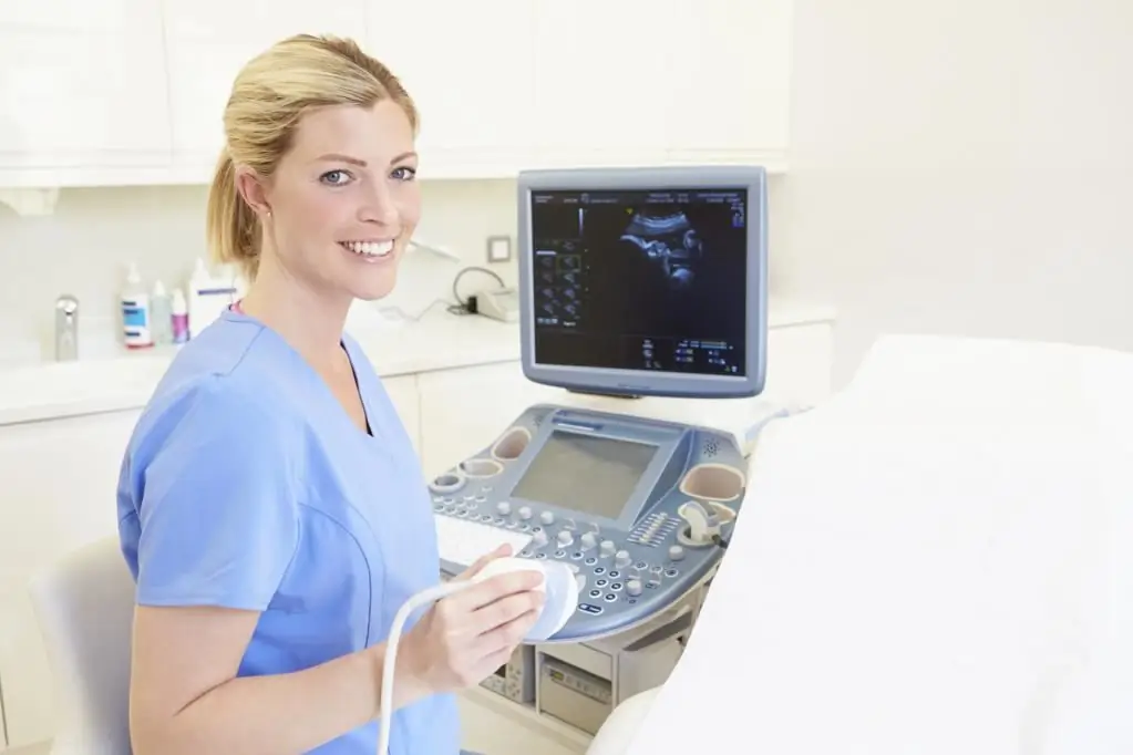
Dopplerometry is performed in the ultrasound room, and its duration depends on a number of factors. For experienced professionals, this takes from 20 to 30 minutes. At the same time, if any pathology is detected, the duration can be increased for obvious reasons.
How is dopplerometry performed during pregnancy? The woman lies on the couch on her back. But sometimes the doctor will ask the woman to turn on her left side, which will be required mainly when the expectant mother is in the third trimester. In this case, the pressure of the enlarged uterus on the inferior vena cava is markedly reduced.
The ultrasound doctor applies a special hypoallergenic and safe gel to the abdomen. Such funds undergo multi-stage testing before use, and only then they are approved for use in relation to pregnant women. The gel has no color and smell, and in its consistency it resembles a thick glue. After that, the doctor picks up a sensor that touches the surface of the skin of the abdomen. An image appears on the screen at this time.

The study allows you to assess the state of the three main blood flow systems, on the basis of whichdopplerometry results:
- fetal PC;
- uteroplacental IPC;
- fetal-placental PPC.
In the study of the BMD, the likelihood of placental insufficiency is determined, while the AUC allows you to identify the severity of this pathology (if any). By PC, you can judge the condition of the child.
Result characteristics
According to the results of the study, one can judge the speed of blood flow in the body of the expectant mother. For this, vascular resistance indices (VR) are determined:
- Resistance Index (RI or IR).
- Ripple index (PI or PI).
- Systole-diastolic ratio (SDR).
RI should be understood as the ratio of the difference between the maximum and minimum blood flow velocities to the maximum value of the velocity in the compression phase. Its formula is: IR=(S-D)/S, where C is the maximum blood flow velocity in the systole phase, and D is the same, only in the diastole phase. The calculation is carried out over several cardiac cycles, and then the average value is determined.
PI is already a slightly different attitude: the same speeds, only to the average blood flow speed. Here the formula is slightly different: PI \u003d (S-D) / M, where M is the average indicator of blood flow velocity.
As for LMS, this abbreviation of dopplerometry during pregnancy hides the ratio of the maximum speed in the systole phase to the minimum rate in the diastole period. Here the formula is simpler: LMS=S-D.
Impaired blood flow
Dopplerometry allows you to diagnose anypathology of fetal development, even in early pregnancy. At the same time, doctors divide them into several degrees depending on the severity of the manifestation:
- IA or IB.
- II degree.
- III degree.
IPC violations are class IA. There are no serious disturbances in the blood circulation of the child, as well as signs of intrauterine growth retardation or fetal development.

Changes in the PPC already apply to class IB. Tellingly, the picture here is the reverse of that presented above. In other words, there are no pathologies in relation to the IPC, and the violations concern only the communication of the blood vessels of the fetus and mother. Just in this case, there may be signs of intrauterine growth retardation and development of the child.
If, during the decoding of Doppler ultrasound during pregnancy, the second degree of severity of the pathology is detected, this indicates the presence of various disorders that affect almost the entire circulatory system of the female body and the fetus. Changes are taking place not only in the IPC, but also in the APC. Although this state of affairs is more serious, the child's life is not yet in danger.
The third degree of blood flow disorder indicates a severe blood flow disorder in a child. As a result, the intake of essential trace elements is significantly reduced, if no measures are taken, this leads to the death of the fetus. Therefore, with such a diagnosis, emergency medical care is needed.
Regardless of the degree of pathological changes in blood flow, a pregnant woman is prescribed the necessary and special coursetreatment. In addition, you will need to go through dopplerometry again to observe the course of changes in dynamics.
Normal indicators
The speed of blood flow is directly dependent on the duration of the child's gestation. Therefore, it is important here to correctly determine the actual pregnancy, otherwise the reliability of the results of the study is called into question. In this regard, only a doctor and no one else should deal with the interpretation of the results. However, only a specialist should also be involved in determining the actual gestational age. In the article you can find a table that reflects the indicators of the norm of dopplerometry during pregnancy by week.

If the results of the conducted studies do not show deviations from the norm, then the development of the child proceeds without complications, all the necessary nutrients are supplied to him in an uninterrupted mode. However, sometimes there may be slight deviations during Doppler. You should not be afraid of this, since all this is easily corrected if necessary.
As a conclusion
From all this we can draw the only correct conclusion: dopplerometry is a fairly reliable and informative diagnosis that allows timely detection of pathological changes in the circulatory system of the female body and fetus. This allows you to make a forecast of the further course of pregnancy and assess the severity of the pathology. Depending on this, it is already necessary to proceed to the necessary measures.
During Doppler withpregnancy, it is also possible to detect hypofunction of the corpus luteum and identify other pathological changes that pose a direct threat to the mother and child. For this reason, you should not ignore all the recommendations of the doctor who leads the pregnancy. Only in this way can many consequences be avoided, and a he althy and full-fledged baby will be born to the delight of parents!
Recommended:
When is the second screening done? Terms, norms, decoding

Examination of a woman's body during pregnancy is mandatory. The performed medical research procedures allow you to monitor the development of the fetus in the womb and, if necessary, correct possible deviations from the norm. This makes it possible to carry a fully developed baby and prevent miscarriage
Norm for screening ultrasound of the 1st trimester. Screening of the 1st trimester: terms, norms for ultrasound, ultrasound interpretation

Why is 1st trimester perinatal screening done? What indicators can be checked by ultrasound in the period of 10-14 weeks?
Weight during pregnancy: norms and deviations. How not to gain weight during pregnancy

What should be the weight during pregnancy? It interests every mother. Many people worry not only about the full development of the baby in the womb, but also about their own figure. Why it is so important to eat right, and what a deficiency or excess weight gain can lead to when carrying crumbs, we will consider in the article
Progesterone norms by week of pregnancy: indicators, causes of deviations at different periods

There are certain norms, in case of deviation from which the probability of developing a successful pregnancy can be extremely small. What are the norms of progesterone by weeks of pregnancy, starting from the moment of conception? Why is this hormone so important for the female body, and what to do if its levels are low? More on this in the article below
Should I do an ultrasound in early pregnancy? Pregnancy on ultrasound in early pregnancy (photo)

Ultrasound came into medicine about 50 years ago. Then this method was used only in exceptional cases. Now, ultrasound machines are in every medical institution. They are used to diagnose the patient's condition, to exclude incorrect diagnoses. Gynecologists also send the patient for ultrasound in early pregnancy

