2026 Author: Priscilla Miln | [email protected]. Last modified: 2025-01-22 17:55:24
When a woman has a serious delay in critical days, the gynecologist sends the patient for ultrasound diagnostics to confirm or deny the presence of pregnancy. First of all, the doctor looks at the contents of the uterus, whether there is a fertilized egg in it.
Why having a fertilized egg is important
A fetal egg found on ultrasound in the uterine cavity is the first confirmation of a he althy uterine pregnancy. At the same time, the thoroughly studied sizes of the fetal egg by weeks make it possible to find out the exact terms of pregnancy, as well as to predict the further course of pregnancy.

From the beginning to the middle of the first trimester, the fertilized egg is one of the main indicators of the good development of the embryo. Since the size of the fetal egg grows by weeks of pregnancy, its size and filling can indicate a successful pregnancy, possible problems, and even a missed pregnancy.
How is the presence of a gestational sac determined
A gynecologist can onlysuggest the presence of a fetal egg in the uterus, based on an increase in the size of the organ. Namely, the doctor can see the fetal egg only with the help of an ultrasound machine.
As a rule, transvaginal ultrasound is performed in the early stages, and it gives the most accurate results, since with this method of diagnosis, the ultrasound can get as close as possible to the object under study.

Fetal egg - what is it?
A fertilized egg is an accumulation of a mass of cells resulting from the fusion of an egg and sperm and the further division of a fertilized egg.
The shape of the mass of cells can be round or oval, but cases of deformation are not excluded. As a rule, diagnosticians look at unusual forms more carefully; in such situations, more frequent observations of the development of the embryo are not excluded. But it’s not worth talking about any problems due to the unusual shape of the egg, since the matter may be in the tone caused by the ultrasound machine itself. By removing the apparatus for a while or by reducing the pressure, the ultrasound specialist can see that the form has changed and returned to normal.
How a fertilized egg appears
A mass of cells travels for some time through the fallopian tube, heading towards the uterus and the place of their future implantation. A week after fertilization has occurred, the fetal egg is attached to any wall of the uterus convenient for this, using the villi located on the outer shell of the egg, destroying a micro-part of the uterine mucosa and vascular walls during implantation. All the time of travel and formation of the fetal egg of the cellfeed on substances from the egg, after which the nutrients will begin to come from the placenta.
At 3 weeks from conception, the size of the fetal egg seriously increases, since a “baby place”, in other words, the placenta, begins to grow from a set of cells embedded in the wall of the uterus. In it, the fetus will live, eat and develop until birth.

The size of the fetal egg will increase again at 5 weeks of pregnancy. At this time, the embryo can already be seen inside the egg. It is worth noting that if at this time the uzist did not see the embryo in the fetal egg, then there is no talk of a missed pregnancy yet and cannot be, since the discrepancy in the timing of the growth of the fetal egg is quite large and can reach two weeks.
The thing is that it is impossible to determine the exact timing of pregnancy during natural conception due to the fact that a woman can ovulate on different days of the cycle, fertilization can also be delayed, attachment can be faster or slower. Therefore, the gestational age is set on the basis of the beginning of the last critical days, which is an obstetric period, and not an embryonic one, and if an embryo is not visible inside the egg at the 5th week of pregnancy, the ultrasound is repeated again after two weeks. Most often, on a second ultrasound, the embryo is already visible.
Weekly sizes
It is not necessary that the size of the fetal egg by week coincide exactly with the standards. Possible error reaches two weeks. In some cases, for example, with late ovulation, the error may be even greater, fet althe egg may have a larger or smaller diameter and this will be the norm, but only if the embryo develops normally.

Here are the sizes of the fetal egg by week on ultrasound:
- Until the 5th week of pregnancy, the fetal egg is very small, by the end of the fifth week it reaches 18 millimeters, and the volume is 2187 millimeters cubed, but by the fourth week it has a diameter of only 7 millimeters. If the egg has a small diameter, then this also indicates a short period that has elapsed since conception.
- Already in the 6th week, the size reaches 22 millimeters.
- At 7 weeks, the size of the ovum is already 24 millimeters.
- In the following weeks, the growth of the ovum is spasmodic, at the eighth week of pregnancy it will be already 30 millimeters, in the future, the egg will grow by an average of 6-8 millimeters weekly.
- By week 13, the diameter will already reach 65 millimeters, and the volume will be 131,070 millimeters cubed.
Growth of the ovum
The size of the fetal egg by week also gives an idea of what size embryo is hidden in the egg. Each week, the embryo develops as rapidly as its house, while the size of the embryo and egg correspond to:
- At 5 weeks, the coccyx-parietal size is 3 millimeters.
- At 6 weeks already 6 millimeters.
- In the 7th week it grows to 10 millimeters.
- At 8 weeks, not only ktr is estimated, but also the biparietal size, namely the estimated width of the head of the embryo, ktr at this time is 16millimeters, and BPR is already 6.
- From week 9 to 13, the fetus grows an average of 10-13 millimeters per week, and by the end of the first trimester, its growth reaches 66 millimeters. The width of the head also grows all this time, at 9 weeks - 8.5 millimeters, at 10 - 11, at 11 - 15 millimeters, at 12 - 20 and at 13 it already reaches 24 millimeters.

It is also worth paying attention to the fact that in the weeks between the main screenings, the size of the egg and the indicators of the embryo may not be so perfectly adjusted, therefore, for screening demonstration studies, weeks are allocated when the fetus approaches the average in all its indicators, in the first trimester, for example, this is 11-14 weeks of pregnancy. Prior to the first screening, at about the 9th week of pregnancy, the uzist and gynecologist check the ratio of the size and the presence of a heartbeat in the fetus, according to these data, adjustments can be made to the estimated gestational age.
How many fertilized eggs can there be
Depending on how many eggs are fertilized at the same time or in a short period of time, one or more fertilized eggs appear.
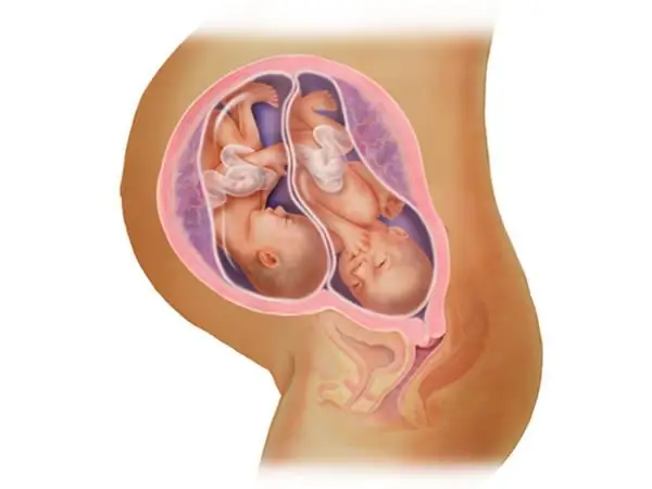
As a rule, if we are talking about twins, that is, about the fertilization of one egg in which two embryos were born, then the fetal egg is one and it is divided into two parts closer to the moment of attachment to the uterine wall or not divided at all. In all other cases, in a multiple pregnancy, there will be as many fetal eggs as there are fertilized eggs, that is, two or more. In conditions of multiplepregnancy, the size of the fetal egg by week will be slightly different from the standards, since the development of the pregnancy itself is a little more complicated, the distribution of nutrients and space in the uterus is also different.
Fetal egg and artificial insemination
Special attention is worthy of the fact that with the advent of such methods for fertilization as IVF, multiple pregnancies, with the development of several fetal bladders at once, has become much more.
With artificial insemination, everything happens a little differently, since an already fertilized fetal egg with an age known to doctors is inserted into the uterus, because the size of each fetal egg usually exactly corresponds to the embryonic period and may not correspond to the gestational age.

All ultrasound transcripts regarding the size of the fetal egg and embryo should be obtained directly from the ultrasound specialist who performed the diagnosis, as well as from a personal gynecologist, since only they will be able to correctly evaluate absolutely all indicators, taking into account the specific features of pregnancy.
Recommended:
Can an ultrasound not show pregnancy? Fetal size by week of pregnancy

There are times when women find out they're pregnant when they're well into their term. There are quite a few ways to confirm a special situation using the analysis of hCG, various tests. But sometimes, the listed methods do not always carry reliable information. Can an ultrasound not show pregnancy? We'll talk about it in this article
Location of the uterus by week of pregnancy. How the size of the uterus and fetus changes every week

Already from the first week after conception, changes imperceptible to the eye begin to occur in the female body. During the examination, the gynecologist can determine the onset of pregnancy by the increased size and location of the uterus. By weeks of pregnancy, an accurate description is provided only according to the results of an ultrasound examination
Size of the abdomen during pregnancy: abdominal circumference by week, fetal development, photo

The size of the abdomen during pregnancy is measured in order to monitor the course of bearing the baby. It helps to determine how the pregnancy is proceeding, whether there are any pathologies. But do not forget about the individual characteristics of the body
What happens in the 12th week of pregnancy. 12 weeks pregnant: fetal size, baby gender, ultrasound picture

12 weeks pregnant is the final stage of the first trimester. During this time, a little man has already developed from a cell that is visible under a microscope, capable of making some movements
Fetometry of the fetus by week. Fetal size by week

For any future mother, it is necessary to be sure that her baby is developing correctly, without various deviations and disorders. Therefore, after the first ultrasound examination, a pregnant woman learns about such a concept as fetometry of the fetus by weeks. Thanks to this type of ultrasound examination, you can find out the dimensions of the body parts of the fetus, make sure that the gestational age set by the doctors is correct and see possible deviations in the dynamics of the child's development

